• 文献检索
  • 文档翻译
  • 深度研究
  • 学术资讯
  • Suppr Zotero 插件Zotero 插件
  • 邀请有礼
  • 套餐&价格
  • 历史记录
应用&插件
Suppr Zotero 插件Zotero 插件浏览器插件Mac 客户端Windows 客户端微信小程序
定价
高级版会员购买积分包购买API积分包
服务
文献检索文档翻译深度研究API 文档MCP 服务
关于我们
关于 Suppr公司介绍联系我们用户协议隐私条款
关注我们

Suppr 超能文献

核心技术专利:CN118964589B侵权必究
粤ICP备2023148730 号-1Suppr @ 2026

文献检索

告别复杂PubMed语法,用中文像聊天一样搜索,搜遍4000万医学文献。AI智能推荐,让科研检索更轻松。

立即免费搜索

文件翻译

保留排版,准确专业,支持PDF/Word/PPT等文件格式,支持 12+语言互译。

免费翻译文档

深度研究

AI帮你快速写综述,25分钟生成高质量综述,智能提取关键信息,辅助科研写作。

立即免费体验

CERA通过影响肾肌成纤维细胞生成减轻db/db小鼠的肾纤维化

CERA Attenuates Kidney Fibrogenesis in the db/db Mouse by Influencing the Renal Myofibroblast Generation.

作者信息

Fischer Christin, Deininger Natalie, Wolf Gunter, Loeffler Ivonne

机构信息

Department of Internal Medicine III, University Hospital Jena, Am Klinikum 1, D-07747 Jena, Germany.

Institute of Medical Genetics and Applied Genomics, University Tübingen, Calwerstraße 7, D-72076 Tübingen, Germany.

出版信息

J Clin Med. 2018 Jan 30;7(2):15. doi: 10.3390/jcm7020015.

DOI:10.3390/jcm7020015
PMID:29385703
原文链接:https://pmc.ncbi.nlm.nih.gov/articles/PMC5852431/
Abstract

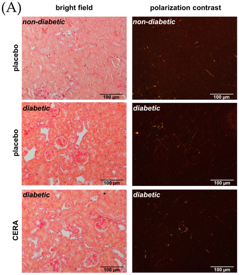
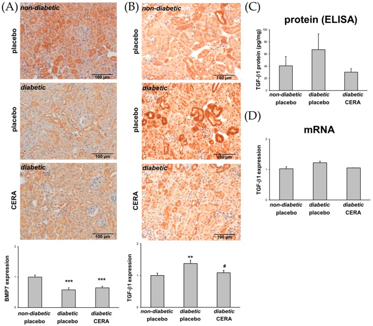
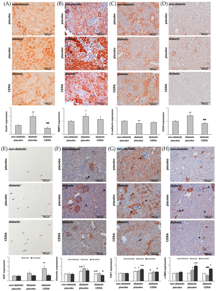
Tubulointerstitial fibrosis (TIF) is a pivotal pathophysiological process in patients with diabetic nephropathy (DN). Multiple profibrotic factors and cell types, including transforming growth factor beta 1 (TGF-β1) and interstitial myofibroblasts, respectively, are responsible for the accumulation of extracellular matrix in the kidney. Matrix-producing myofibroblasts can originate from different sources and different mechanisms are involved in the activation process of the myofibroblasts in the fibrotic kidney. In this study, 16-week-old / mice, a model for type 2 DN, were treated for two weeks with continuous erythropoietin receptor activator (CERA), a synthetic erythropoietin variant with possible non-hematopoietic, tissue-protective effects. Non-diabetic and diabetic mice treated with placebo were used as controls. The effects of CERA on tubulointerstitial fibrosis (TIF) as well as on the generation of the matrix-producing myofibroblasts were evaluated by morphological, immunohistochemical, and molecular biological methods. The placebo-treated diabetic mice showed significant signs of beginning renal TIF (shown by picrosirius red staining; increased connective tissue growth factor (CTGF), fibronectin and collagen I deposition; upregulated KIM1 expression) together with an increased number of interstitial myofibroblasts (shown by different mesenchymal markers), while kidneys from diabetic mice treated with CERA revealed less TIF and fewer myofibroblasts. The mechanisms, in which CERA acts as an anti-fibrotic agent/drug, seem to be multifaceted: first, CERA inhibits the generation of matrix-producing myofibroblasts and second, CERA increases the ability for tissue repair. Many of these CERA effects can be explained by the finding that CERA inhibits the renal expression of the cytokine TGF-β1.

摘要

肾小管间质纤维化(TIF)是糖尿病肾病(DN)患者关键的病理生理过程。多种促纤维化因子和细胞类型,分别包括转化生长因子β1(TGF-β1)和间质肌成纤维细胞,是肾脏细胞外基质积累的原因。产生基质的肌成纤维细胞可源自不同来源,且纤维化肾脏中肌成纤维细胞激活过程涉及不同机制。在本研究中,16周龄的2型糖尿病肾病模型小鼠连续两周接受促红细胞生成素受体激活剂(CERA)治疗,CERA是一种具有可能的非造血、组织保护作用的合成促红细胞生成素变体。用安慰剂治疗的非糖尿病和糖尿病小鼠作为对照。通过形态学、免疫组织化学和分子生物学方法评估CERA对肾小管间质纤维化(TIF)以及对产生基质的肌成纤维细胞生成的影响。用安慰剂治疗的糖尿病小鼠显示出肾脏TIF开始的显著迹象(用天狼星红染色显示;结缔组织生长因子(CTGF)、纤连蛋白和I型胶原沉积增加;KIM1表达上调),同时间质肌成纤维细胞数量增加(用不同间充质标志物显示),而用CERA治疗的糖尿病小鼠的肾脏显示出较少的TIF和较少的肌成纤维细胞。CERA作为抗纤维化药物发挥作用的机制似乎是多方面的:首先,CERA抑制产生基质的肌成纤维细胞的生成;其次,CERA增强组织修复能力。CERA的许多这些作用可以通过CERA抑制细胞因子TGF-β1在肾脏中的表达这一发现来解释。

https://cdn.ncbi.nlm.nih.gov/pmc/blobs/ddcc/5852431/49afa696947e/jcm-07-00015-g003.jpg
https://cdn.ncbi.nlm.nih.gov/pmc/blobs/ddcc/5852431/ab8bd5fd538b/jcm-07-00015-g001a.jpg
https://cdn.ncbi.nlm.nih.gov/pmc/blobs/ddcc/5852431/e62e36e5b9fa/jcm-07-00015-g002.jpg
https://cdn.ncbi.nlm.nih.gov/pmc/blobs/ddcc/5852431/49afa696947e/jcm-07-00015-g003.jpg
https://cdn.ncbi.nlm.nih.gov/pmc/blobs/ddcc/5852431/ab8bd5fd538b/jcm-07-00015-g001a.jpg
https://cdn.ncbi.nlm.nih.gov/pmc/blobs/ddcc/5852431/e62e36e5b9fa/jcm-07-00015-g002.jpg
https://cdn.ncbi.nlm.nih.gov/pmc/blobs/ddcc/5852431/49afa696947e/jcm-07-00015-g003.jpg

相似文献

1
CERA Attenuates Kidney Fibrogenesis in the db/db Mouse by Influencing the Renal Myofibroblast Generation.CERA通过影响肾肌成纤维细胞生成减轻db/db小鼠的肾纤维化
J Clin Med. 2018 Jan 30;7(2):15. doi: 10.3390/jcm7020015.
2
FSP1-specific SMAD2 knockout in renal tubular, endothelial, and interstitial cells reduces fibrosis and epithelial-to-mesenchymal transition in murine STZ-induced diabetic nephropathy.FSP1 特异性 SMAD2 敲除在肾小管、内皮和间质细胞中可减少 STZ 诱导的糖尿病肾病小鼠的纤维化和上皮间质转化。
Cell Tissue Res. 2018 Apr;372(1):115-133. doi: 10.1007/s00441-017-2754-1. Epub 2017 Dec 6.
3
Erythropoietin ameliorates podocyte injury in advanced diabetic nephropathy in the db/db mouse.促红细胞生成素可改善 db/db 糖尿病小鼠晚期糖尿病肾病足细胞损伤。
Am J Physiol Renal Physiol. 2013 Sep 15;305(6):F911-8. doi: 10.1152/ajprenal.00643.2012. Epub 2013 Jul 3.
4
The continuous erythropoietin receptor activator affects different pathways of diabetic renal injury.持续促红细胞生成素受体激活剂影响糖尿病肾损伤的不同途径。
J Am Soc Nephrol. 2007 Jul;18(7):2046-53. doi: 10.1681/ASN.2006070699. Epub 2007 Jun 6.
5
Renal tubulointerstitial fibrosis in OVE26 type 1 diabetic mice.OVE26型1型糖尿病小鼠的肾小管间质纤维化
Nephron Exp Nephrol. 2009;111(1):e11-9. doi: 10.1159/000178763. Epub 2008 Dec 2.
6
Chronic erythropoietin treatment affects different molecular pathways of diabetic cardiomyopathy in mouse.慢性促红细胞生成素治疗影响小鼠糖尿病心肌病的不同分子途径。
Eur J Clin Invest. 2009 Sep;39(9):755-60. doi: 10.1111/j.1365-2362.2009.02165.x. Epub 2009 Jul 14.
7
Increased glomerular and tubular expression of transforming growth factor-beta1, its type II receptor, and activation of the Smad signaling pathway in the db/db mouse.db/db小鼠中转化生长因子-β1及其II型受体的肾小球和肾小管表达增加,以及Smad信号通路的激活。
Am J Pathol. 2001 May;158(5):1653-63. doi: 10.1016/s0002-9440(10)64121-1.
8
Transformation of interstitial fibroblasts and tubulointerstitial fibrosis in diabetic nephropathy.糖尿病肾病中肾间质成纤维细胞的转化与肾小管间质纤维化
Med Electron Microsc. 2002 Jun;35(2):87-95. doi: 10.1007/s007950200011.
9
Pericyte activation accompanied by peritubular capillaries dysfunction and pericyte-to-myofibroblast transition is associated with renal fibrosis in diabetic nephropathy.周细胞激活伴有肾小管周围毛细血管功能障碍以及周细胞向肌成纤维细胞转变,这与糖尿病肾病中的肾纤维化有关。
Kidney Res Clin Pract. 2024 Feb 6. doi: 10.23876/j.krcp.23.099.
10
Connective tissue growth factor expressed in tubular epithelium plays a pivotal role in renal fibrogenesis.在肾小管上皮细胞中表达的结缔组织生长因子在肾纤维化形成过程中起关键作用。
J Am Soc Nephrol. 2005 Jan;16(1):133-43. doi: 10.1681/ASN.2004040339. Epub 2004 Dec 1.

引用本文的文献

1
New Approaches to Diabetic Nephropathy from Bed to Bench.糖尿病肾病从临床到实验室研究的新方法
Biomedicines. 2022 Apr 9;10(4):876. doi: 10.3390/biomedicines10040876.
2
Diabetic fibrosis.糖尿病性纤维化
Biochim Biophys Acta Mol Basis Dis. 2021 Apr 1;1867(4):166044. doi: 10.1016/j.bbadis.2020.166044. Epub 2020 Dec 28.
3
Effects of Allicin on Pathophysiological Mechanisms during the Progression of Nephropathy Associated to Diabetes.大蒜素对糖尿病相关性肾病进展过程中病理生理机制的影响

本文引用的文献

1
Heterozygosity of mitogen-activated protein kinase organizer 1 ameliorates diabetic nephropathy and suppresses epithelial-to-mesenchymal transition-like changes in db/db mice.丝裂原活化蛋白激酶组织器 1 的杂合性改善糖尿病肾病,并抑制 db/db 小鼠的上皮间质转化样改变。
Nephrol Dial Transplant. 2017 Dec 1;32(12):2017-2034. doi: 10.1093/ndt/gfx202.
2
Snail and kidney fibrosis.蜗牛与肾脏纤维化。
Nephrol Dial Transplant. 2017 Feb 1;32(2):224-233. doi: 10.1093/ndt/gfw333.
3
The origin of renal fibroblasts/myofibroblasts and the signals that trigger fibrosis.
Antioxidants (Basel). 2020 Nov 15;9(11):1134. doi: 10.3390/antiox9111134.
4
Recombinant Erythropoietin Provides Protection against Renal Fibrosis in Adenine-Induced Chronic Kidney Disease.重组促红细胞生成素对腺嘌呤诱导的慢性肾脏病肾纤维化的保护作用。
Mediators Inflamm. 2020 Feb 27;2020:8937657. doi: 10.1155/2020/8937657. eCollection 2020.
5
Advances in Understanding the Effects of Erythropoietin on Renal Fibrosis.促红细胞生成素对肾纤维化影响的认识进展
Front Med (Lausanne). 2020 Feb 21;7:47. doi: 10.3389/fmed.2020.00047. eCollection 2020.
6
FoxO1-mediated inhibition of STAT1 alleviates tubulointerstitial fibrosis and tubule apoptosis in diabetic kidney disease.FoxO1 介导的 STAT1 抑制减轻糖尿病肾病中的肾小管间质纤维化和肾小管细胞凋亡。
EBioMedicine. 2019 Oct;48:491-504. doi: 10.1016/j.ebiom.2019.09.002. Epub 2019 Oct 16.
7
Changes in cell fate determine the regenerative and functional capacity of the developing kidney before and after release of obstruction.细胞命运的改变决定了肾脏在解除梗阻前后的再生和功能能力。
Clin Sci (Lond). 2018 Dec 5;132(23):2519-2545. doi: 10.1042/CS20180623. Print 2018 Dec 12.
肾成纤维细胞/肌成纤维细胞的起源以及引发纤维化的信号。
Differentiation. 2016 Sep;92(3):102-107. doi: 10.1016/j.diff.2016.05.008. Epub 2016 Jun 1.
4
A chemical screen to identify inducers of the mitochondrial unfolded protein response in C. elegans.一项用于鉴定秀丽隐杆线虫中线粒体未折叠蛋白反应诱导剂的化学筛选。
Worm. 2015 Oct 2;4(4):e1096490. doi: 10.1080/21624054.2015.1096490. eCollection 2015 Oct-Dec.
5
Pleiotropic and Renoprotective Effects of Erythropoietin Beta on Experimental Diabetic Nephropathy Model.促红细胞生成素β对实验性糖尿病肾病模型的多效性及肾脏保护作用
Nephron. 2016;132(4):292-300. doi: 10.1159/000444649. Epub 2016 Mar 4.
6
MicroRNA-130b improves renal tubulointerstitial fibrosis via repression of Snail-induced epithelial-mesenchymal transition in diabetic nephropathy.微小RNA-130b通过抑制Snail诱导的糖尿病肾病肾小管间质纤维化中的上皮-间质转化来改善病情。
Sci Rep. 2016 Feb 3;6:20475. doi: 10.1038/srep20475.
7
Epithelial-to-mesenchymal transition of tubular epithelial cells in renal fibrosis: a new twist on an old tale.肾小管上皮细胞的上皮-间充质转化在肾纤维化中的作用:旧故事的新转折。
Kidney Int. 2016 Feb;89(2):263-6. doi: 10.1016/j.kint.2015.12.025.
8
Matrix Metalloproteinase-2 Knockout and Heterozygote Mice Are Protected from Hydronephrosis and Kidney Fibrosis after Unilateral Ureteral Obstruction.基质金属蛋白酶-2基因敲除小鼠和杂合子小鼠在单侧输尿管梗阻后可免受肾积水和肾纤维化的影响。
PLoS One. 2015 Dec 16;10(12):e0143390. doi: 10.1371/journal.pone.0143390. eCollection 2015.
9
Epithelial-to-Mesenchymal Transition in Diabetic Nephropathy: Fact or Fiction?糖尿病肾病中的上皮-间充质转化:事实还是虚构?
Cells. 2015 Oct 9;4(4):631-52. doi: 10.3390/cells4040631.
10
Smad1/5/8 are myogenic regulators of murine and human mesoangioblasts.Smad1/5/8是小鼠和人类中胚层血管母细胞的生肌调节因子。
J Mol Cell Biol. 2016 Feb;8(1):73-87. doi: 10.1093/jmcb/mjv059. Epub 2015 Oct 8.