Dittmann Isabel L, Zauchner Thomas, Nevard Lucy M, Telford Maximilian J, Egger Bernhard
Research unit Evolutionary Developmental Biology, Institute of Zoology, University of Innsbruck, Technikerstr. 25, Innsbruck, 6020, Austria.
Department of Genetics, Evolution and Environment, University College London, Darwin Building, Gower Street, London, WC1E 6BT, United Kingdom.
J Morphol. 2018 May;279(5):589-597. doi: 10.1002/jmor.20794. Epub 2018 Feb 1.
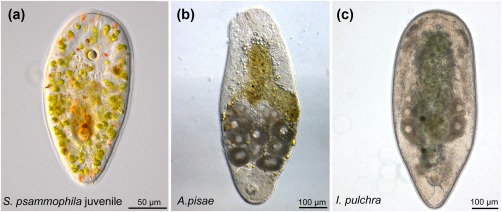
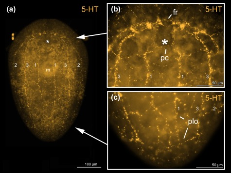
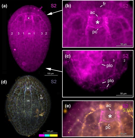
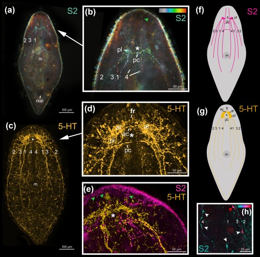
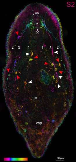
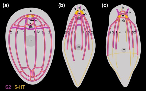
Acoel worms are simple, often microscopic animals with direct development, a multiciliated epidermis, a statocyst, and a digestive parenchyma instead of a gut epithelium. Morphological characters of acoels have been notoriously difficult to interpret due to their relative scarcity. The nervous system is one of the most accessible and widely used comparative features in acoels, which have a so-called commissural brain without capsule and several major longitudinal neurite bundles. Here, we use the selective binding properties of a neuropeptide antibody raised in echinoderms (SALMFamide2, or S2), and a commercial antibody against serotonin (5-HT) to provide additional characters of the acoel nervous system. We have prepared whole-mount immunofluorescent stainings of three acoel species: Symsagittifera psammophila (Convolutidae), Aphanostoma pisae, and the model acoel Isodiametra pulchra (both Isodiametridae). The commissural brain of all three acoels is delimited anteriorly by the ventral anterior commissure, and posteriorly by the dorsal posterior commissure. The dorsal anterior commissure is situated between the ventral anterior commissure and the dorsal posterior commissure, while the statocyst lies between dorsal anterior and dorsal posterior commissure. S2 and serotonin do not co-localise, and they follow similar patterns to each other within an animal. In particular, S2, but not 5-HT, stains a prominent commissure posterior to the main (dorsal) posterior commissure. We have for the first time observed a closed posterior loop of the main neurite bundles in S. psammophila for both the amidergic and the serotonergic nervous system. In I. pulchra, the lateral neurite bundles also form a posterior loop in our serotonergic nervous system stainings.
无肠目蠕虫是简单的动物,通常体型微小,具有直接发育方式、多纤毛表皮、平衡囊以及由消化实质组织而非肠上皮构成的消化系统。由于无肠目蠕虫相对稀少,其形态特征一直难以解释。神经系统是无肠目蠕虫中最易于观察且被广泛用于比较研究的特征之一,它们具有一个所谓的无包膜连合脑以及几条主要的纵向神经突束。在此,我们利用在棘皮动物中产生的一种神经肽抗体(SALMFamide2,或S2)以及一种针对血清素(5 - HT)的商业抗体的选择性结合特性,来提供无肠目蠕虫神经系统的更多特征。我们制备了三种无肠目物种的整体免疫荧光染色标本:沙栖对称涡虫(Convolutidae科)、豌豆无吻涡虫以及模式无肠目物种秀丽等径涡虫(均为Isodiametridae科)。所有这三种无肠目蠕虫的连合脑在前方由腹侧前连合界定,后方由背侧后连合界定。背侧前连合位于腹侧前连合和背侧后连合之间,而平衡囊则位于背侧前连合和背侧后连合之间。S2和血清素不共定位,并且它们在动物体内呈现相似的模式。特别是,S2而非5 - HT,标记了一条位于主要(背侧)后连合后方的显著连合。我们首次在沙栖对称涡虫中观察到了主神经突束的一个闭合后环,涉及酰胺能和血清素能神经系统。在秀丽等径涡虫中,在我们的血清素能神经系统染色中,外侧神经突束也形成了一个后环。