• 文献检索
  • 文档翻译
  • 深度研究
  • 学术资讯
  • Suppr Zotero 插件Zotero 插件
  • 邀请有礼
  • 套餐&价格
  • 历史记录
应用&插件
Suppr Zotero 插件Zotero 插件浏览器插件Mac 客户端Windows 客户端微信小程序
定价
高级版会员购买积分包购买API积分包
服务
文献检索文档翻译深度研究API 文档MCP 服务
关于我们
关于 Suppr公司介绍联系我们用户协议隐私条款
关注我们

Suppr 超能文献

核心技术专利:CN118964589B侵权必究
粤ICP备2023148730 号-1Suppr @ 2026

文献检索

告别复杂PubMed语法,用中文像聊天一样搜索,搜遍4000万医学文献。AI智能推荐,让科研检索更轻松。

立即免费搜索

文件翻译

保留排版,准确专业,支持PDF/Word/PPT等文件格式,支持 12+语言互译。

免费翻译文档

深度研究

AI帮你快速写综述,25分钟生成高质量综述,智能提取关键信息,辅助科研写作。

立即免费体验

体外冲击波疗法以低能量通量密度抑制动物模型中的增生性瘢痕形成。

Extracorporeal shock wave therapy with low-energy flux density inhibits hypertrophic scar formation in an animal model.

机构信息

Burns and Plastic Reconstruction Unit, The First Hospital of Jilin University, Changchun, Jilin 130021, P.R. China.

出版信息

Int J Mol Med. 2018 Apr;41(4):1931-1938. doi: 10.3892/ijmm.2018.3434. Epub 2018 Jan 29.

DOI:10.3892/ijmm.2018.3434
PMID:29393337
原文链接:https://pmc.ncbi.nlm.nih.gov/articles/PMC5810209/
Abstract

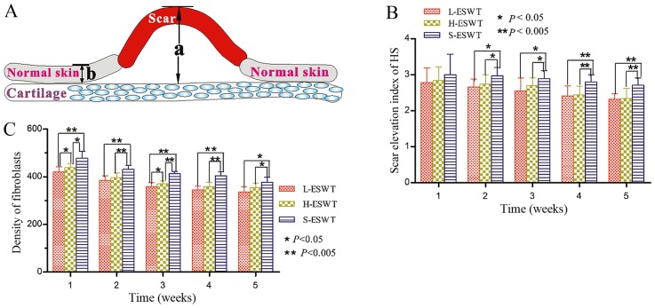
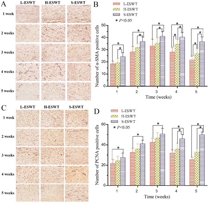
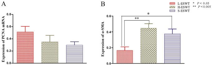
Hypertrophic scar is characterized by excessive deposits of collagen during skin wound healing, which could become a challenge to clinicians. This study assessed the effects of the extracorporeal shock wave therapy (ESWT) on hypertrophic scar formation and the underlying gene regu-lation. A rabbit ear hypertrophic scar model was generated and randomly divided into three groups: L-ESWT group to receive L-ESWT (energy flux density of 0.1 mJ/mm2), H-ESWT (energy flux density of 0.2 mJ/mm2) and sham ESWT group (S-ESWT). Hypertrophic scar tissues were then collected and stained with hematoxylin and eosin (H&E) and Masson's trichrome staining, respectively, to assess scar elevation index (SEI), fibroblast density and collagen fiber arrangement. Expression of cell proliferation marker proliferating cell nuclear antigen (PCNA) and α-smooth muscle actin (α-SMA) were assessed using RT-PCR and immunohistochemistry in hypertrophic scar tissues. H&E staining sections showed significant reduction of SEI and fibroblast density in both ESWT treatment groups compared to S-ESWT, but there was no dramatic difference between L-ESWT and H-ESWT groups. Masson's trichrome staining showed that collagen fibers were more slender and broader and oriented in parallel to skin surface after administration of ESWT compared to control tissues. At the gene level, PCNA‑positive fibroblasts and α-SMA-positive myofibroblasts were significantly decreased after L-ESWT or H-ESWT compared to the controls. Furthermore, there was no significant difference in expression of PCNA mRNA between L-ESWT or H-ESWT and S-ESWT, whereas expression of α-SMA mRNA significantly decreased in L-ESWT compared to that of H-ESWT and S-ESWT (P=0.002 and P=0.030, respectively). In conclusion, L-ESWT could be effective on suppression of hypertrophic scar formation by inhibition of scar elevation index and fibroblast density as well as α-SMA expression in hypertrophic scar tissues of the rabbit model.

摘要

增生性瘢痕的特征是皮肤创伤愈合过程中胶原过度沉积,这给临床医生带来了挑战。本研究评估了体外冲击波疗法(ESWT)对增生性瘢痕形成及潜在基因调控的影响。建立兔耳增生性瘢痕模型,并随机分为三组:低能冲击波组(L-ESWT,能量通量密度为 0.1mJ/mm2)、高能冲击波组(H-ESWT,能量通量密度为 0.2mJ/mm2)和假冲击波组(S-ESWT)。然后采集增生性瘢痕组织,分别用苏木精和伊红(H&E)和马松三色染色法染色,评估瘢痕隆起指数(SEI)、成纤维细胞密度和胶原纤维排列。采用 RT-PCR 和免疫组织化学法检测增生性瘢痕组织中细胞增殖标志物增殖细胞核抗原(PCNA)和α-平滑肌肌动蛋白(α-SMA)的表达。H&E 染色切片显示,与 S-ESWT 相比,两种 ESWT 治疗组的 SEI 和成纤维细胞密度均显著降低,但 L-ESWT 组与 H-ESWT 组之间无显著差异。马松三色染色显示,与对照组织相比,ESWT 治疗后胶原纤维更纤细、更宽,并与皮肤表面平行排列。在基因水平上,与对照组相比,L-ESWT 或 H-ESWT 后 PCNA 阳性成纤维细胞和α-SMA 阳性肌成纤维细胞的数量明显减少。此外,L-ESWT 或 H-ESWT 与 S-ESWT 之间 PCNA mRNA 的表达无显著差异,而 L-ESWT 组α-SMA mRNA 的表达明显低于 H-ESWT 组和 S-ESWT 组(P=0.002 和 P=0.030)。结论,L-ESWT 可通过抑制兔模型增生性瘢痕组织中的 SEI 和成纤维细胞密度以及α-SMA 表达来有效抑制增生性瘢痕形成。

https://cdn.ncbi.nlm.nih.gov/pmc/blobs/ed3e/5810209/914abe41f45a/IJMM-41-04-1931-g04.jpg
https://cdn.ncbi.nlm.nih.gov/pmc/blobs/ed3e/5810209/d767c69d892a/IJMM-41-04-1931-g00.jpg
https://cdn.ncbi.nlm.nih.gov/pmc/blobs/ed3e/5810209/a041693530bf/IJMM-41-04-1931-g01.jpg
https://cdn.ncbi.nlm.nih.gov/pmc/blobs/ed3e/5810209/df28c21e4b8b/IJMM-41-04-1931-g02.jpg
https://cdn.ncbi.nlm.nih.gov/pmc/blobs/ed3e/5810209/c3e00e9e47f1/IJMM-41-04-1931-g03.jpg
https://cdn.ncbi.nlm.nih.gov/pmc/blobs/ed3e/5810209/914abe41f45a/IJMM-41-04-1931-g04.jpg

相似文献

1
Extracorporeal shock wave therapy with low-energy flux density inhibits hypertrophic scar formation in an animal model.体外冲击波疗法以低能量通量密度抑制动物模型中的增生性瘢痕形成。
Int J Mol Med. 2018 Apr;41(4):1931-1938. doi: 10.3892/ijmm.2018.3434. Epub 2018 Jan 29.
2
Extracorporeal Shock Wave Therapy Alters the Expression of Fibrosis-Related Molecules in Fibroblast Derived from Human Hypertrophic Scar.体外冲击波疗法改变人增生性瘢痕成纤维细胞中纤维化相关分子的表达。
Int J Mol Sci. 2018 Jan 2;19(1):124. doi: 10.3390/ijms19010124.
3
Adipose tissue-derived stem cells suppress hypertrophic scar fibrosis via the p38/MAPK signaling pathway.脂肪组织来源的干细胞通过p38/丝裂原活化蛋白激酶信号通路抑制增生性瘢痕纤维化。
Stem Cell Res Ther. 2016 Aug 2;7(1):102. doi: 10.1186/s13287-016-0356-6.
4
Lower energy radial shock wave therapy improves characteristics of hypertrophic scar in a rabbit ear model.低能量径向冲击波疗法改善兔耳模型中增生性瘢痕的特征。
Exp Ther Med. 2018 Jan;15(1):933-939. doi: 10.3892/etm.2017.5441. Epub 2017 Nov 6.
5
Effectiveness of fractional carbon dioxide laser combined with botulinum toxin type A in a rabbit ear model with the underlying mechanism.分比二氧化碳激光联合肉毒毒素 A 治疗兔耳模型的有效性及其潜在机制。
J Cosmet Dermatol. 2023 Aug;22(8):2225-2232. doi: 10.1111/jocd.15703. Epub 2023 Mar 13.
6
Cryptotanshinone downregulates the profibrotic activities of hypertrophic scar fibroblasts and accelerates wound healing: A potential therapy for the reduction of skin scarring.隐丹参酮下调增生性瘢痕成纤维细胞的促纤维化活性并加速伤口愈合:减少皮肤瘢痕形成的一种潜在治疗方法。
Biomed Pharmacother. 2016 May;80:80-86. doi: 10.1016/j.biopha.2016.03.006. Epub 2016 Mar 17.
7
Anti-inflammatory cytokine TSG-6 inhibits hypertrophic scar formation in a rabbit ear model.抗炎细胞因子TSG-6抑制兔耳模型中的增生性瘢痕形成。
Eur J Pharmacol. 2015 Mar 15;751:42-9. doi: 10.1016/j.ejphar.2015.01.040. Epub 2015 Feb 3.
8
Effect of extracorporeal shock wave therapy for burn scar regeneration: A prospective, randomized, double-blinded study.体外冲击波治疗烧伤后再生的效果:一项前瞻性、随机、双盲研究。
Burns. 2021 Jun;47(4):821-827. doi: 10.1016/j.burns.2020.08.009. Epub 2020 Aug 29.
9
[Effect of tetrandine on gene expression of collagen type I, collagen type III and TGF-beta1 in scar tissue's of rabbits ear].汉防己甲素对兔耳瘢痕组织中Ⅰ型胶原、Ⅲ型胶原及转化生长因子β1基因表达的影响
Zhonghua Zheng Xing Wai Ke Za Zhi. 2013 Nov;29(6):406-12.
10
Comparison of Enalapril, Candesartan and Intralesional Triamcinolone in Reducing Hypertrophic Scar Development: An Experimental Study.依那普利、坎地沙坦与曲安奈德局部注射在减少肥厚性瘢痕形成中的比较:一项实验研究
Aesthetic Plast Surg. 2018 Apr;42(2):352-361. doi: 10.1007/s00266-018-1073-6. Epub 2018 Jan 18.

引用本文的文献

1
Chrysin inhibits hypertrophic scar formation through TGF-β/Smad signaling pathways.白杨素通过TGF-β/Smad信号通路抑制增生性瘢痕形成。
J Mol Histol. 2025 Sep 6;56(5):298. doi: 10.1007/s10735-025-10576-3.
2
Mechanisms of extracorporeal shock wave therapy in burn scar repair: Advances in immunoactivation-based scar remodeling research.体外冲击波疗法在烧伤瘢痕修复中的作用机制:基于免疫激活的瘢痕重塑研究进展
Regen Ther. 2025 Jun 12;30:182-194. doi: 10.1016/j.reth.2025.05.010. eCollection 2025 Dec.
3
Effect of Extracorporeal Shock Wave Therapy on Post-Laminectomy Lumbar Epidural Fibrosis.

本文引用的文献

1
[Advances in the research of mechanism of enhancement of wound healing with extracorporeal shock wave therapy].[体外冲击波疗法促进伤口愈合的机制研究进展]
Zhonghua Shao Shang Za Zhi. 2015 Aug;31(4):315-7.
2
Lateral epicondylitis: This is still a main indication for extracorporeal shockwave therapy.外侧肱骨上髁炎:这仍然是体外冲击波疗法的主要适应证。
Int J Surg. 2015 Dec;24(Pt B):165-70. doi: 10.1016/j.ijsu.2015.09.034. Epub 2015 Oct 9.
3
Extracorporeal Shock Wave Therapy: An Emerging Treatment Modality for Retracting Scars of the Hands.
体外冲击波疗法对椎板切除术后腰椎硬膜外纤维化的影响。
Ann Rehabil Med. 2025 Apr;49(2):81-90. doi: 10.5535/arm.240118. Epub 2025 Apr 30.
4
Systematic Review on Working Mechanisms of Signaling Pathways in Fibrosis During Shockwave Therapy.冲击波治疗休克期间纤维化信号通路作用机制的系统评价
Int J Mol Sci. 2024 Oct 31;25(21):11729. doi: 10.3390/ijms252111729.
5
The Role of Extracorporeal Shock Wave Therapy in Keloids and Hypertrophic Scars: A Systematic Review.体外冲击波疗法在瘢痕疙瘩和增生性瘢痕中的作用:一项系统评价
Cureus. 2024 Oct 19;16(10):e71869. doi: 10.7759/cureus.71869. eCollection 2024 Oct.
6
A Preliminary Study on Quantitative Analysis of Collagen and Apoptosis Related Protein on 1064 nm Laser-Induced Skin Injury.1064nm激光致皮肤损伤中胶原蛋白及凋亡相关蛋白定量分析的初步研究
Biology (Basel). 2024 Mar 27;13(4):217. doi: 10.3390/biology13040217.
7
Extracorporeal Shockwave Treatment as Additional Therapy in Patients with Post-Stroke Spasticity of Upper Limb-A Narrative Review.体外冲击波治疗作为中风后上肢痉挛患者的辅助治疗——一项叙述性综述
J Clin Med. 2024 Mar 30;13(7):2017. doi: 10.3390/jcm13072017.
8
Pain-Relieving Effects of Shockwave Therapy for Ledderhose Disease: An Ultrasound-Based Study of an Unusual Bilateral Case.体外冲击波疗法治疗足底纤维瘤病的止痛效果:基于超声的罕见双侧病例研究
Life (Basel). 2024 Jan 24;14(2):169. doi: 10.3390/life14020169.
9
Targeting Signalling Pathways in Chronic Wound Healing.靶向慢性伤口愈合中的信号通路。
Int J Mol Sci. 2023 Dec 19;25(1):50. doi: 10.3390/ijms25010050.
10
Possible mechanism of static progressive stretching combined with extracorporeal shock wave therapy in reducing knee joint contracture in rats based on MAPK/ERK pathway.基于 MAPK/ERK 通路探讨静态渐进牵伸联合体外冲击波疗法减轻大鼠膝关节挛缩的可能机制。
Biomol Biomed. 2023 Mar 16;23(2):277-286. doi: 10.17305/bjbms.2022.8152.
体外冲击波疗法:一种用于手部瘢痕退缩的新兴治疗方式。
Ultrasound Med Biol. 2016 Jan;42(1):185-95. doi: 10.1016/j.ultrasmedbio.2015.07.028. Epub 2015 Oct 9.
4
Extracorporeal shockwave therapy in diabetic foot ulcers.体外冲击波疗法治疗糖尿病足溃疡。
Int J Surg. 2015 Dec;24(Pt B):207-9. doi: 10.1016/j.ijsu.2015.06.024. Epub 2015 Jun 12.
5
In vivo biological response to extracorporeal shockwave therapy in human tendinopathy.人体肌腱病中体外冲击波疗法的体内生物学反应
Eur Cell Mater. 2015 May 15;29:268-80; discussion 280. doi: 10.22203/ecm.v029a20.
6
Assessment of the Effectiveness of Extracorporeal Shock Wave Therapy (ESWT) For Soft Tissue Injuries (ASSERT): An Online Database Protocol.体外冲击波疗法(ESWT)治疗软组织损伤有效性评估(ASSERT):一项在线数据库方案
Transl Med UniSa. 2014 Apr 8;10:46-51. eCollection 2014 Sep.
7
Extracorporeal shock wave therapy as an adjunct wound treatment: a systematic review of the literature.体外冲击波疗法作为一种辅助伤口治疗方法:文献系统综述
Ostomy Wound Manage. 2014 Jul;60(7):26-39.
8
Induction of galectin-1 by TGF-β1 accelerates fibrosis through enhancing nuclear retention of Smad2.转化生长因子-β1 诱导半乳糖凝集素-1 通过增强 Smad2 的核滞留加速纤维化。
Exp Cell Res. 2014 Aug 1;326(1):125-35. doi: 10.1016/j.yexcr.2014.06.001. Epub 2014 Jun 11.
9
Is extracorporeal shock wave therapy clinical efficacy for relief of chronic, recalcitrant plantar fasciitis? A systematic review and meta-analysis of randomized placebo or active-treatment controlled trials.体外冲击波疗法治疗慢性、难治性足底筋膜炎的临床疗效如何?一项随机安慰剂或阳性药物对照试验的系统评价和荟萃分析。
Arch Phys Med Rehabil. 2014 Aug;95(8):1585-93. doi: 10.1016/j.apmr.2014.01.033. Epub 2014 Mar 21.
10
Extracorporeal shock wave lithotripsy: An opinion on its future.体外冲击波碎石术:对其未来的看法。
Indian J Urol. 2014 Jan;30(1):73-9. doi: 10.4103/0970-1591.124211.