Kreltszheim Michael A S, Brown Nick I, Lee Joseph C
Department of Medical Imaging, Gold Coast University Hospital, Southport QLD 4215, Australia.
Department of Medical Imaging, The Prince Charles Hospital, Chermside QLD 4032, Australia.
World J Nucl Med. 2018 Jan-Mar;17(1):59-61. doi: 10.4103/wjnm.WJNM_3_17.
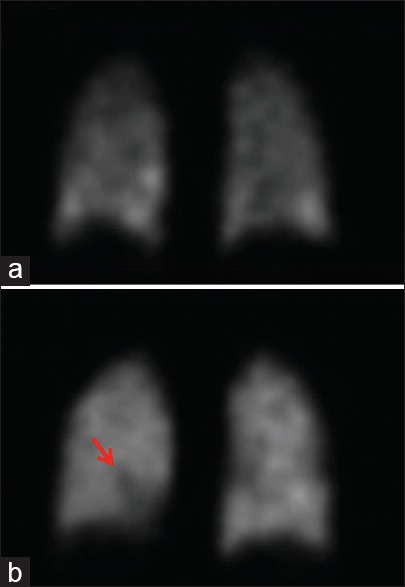
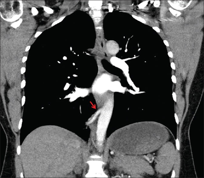
We present a 46-year-old female with pleuritic chest pain on a background of pulmonary embolism diagnosed on a single-photon emission computed tomography (SPECT) ventilation-perfusion (V/Q) imaging 3 years earlier. A SPECT V/Q scan detected a mismatched perfusion defect in the posterior basal segment of the right lower lobe, essentially unchanged from a defect identified 3 years earlier. Given the atypical finding, the patient went on to have a computed tomographic pulmonary angiogram. It revealed an intralobar bronchopulmonary sequestration as the cause of the right lower lobe mismatched perfusion defect. With growing awareness of radiation safety, the number of V/Q imaging studies being undertaken to investigate suspected pulmonary emboli, especially in young female patients, has increased. This case report serves as a timely reminder of the potential pitfalls associated with V/Q scan image interpretation.
我们报告一例46岁女性,有胸膜炎性胸痛,3年前经单光子发射计算机断层扫描(SPECT)通气灌注(V/Q)显像诊断为肺栓塞。SPECT V/Q扫描在右下叶后基底段发现一个不匹配的灌注缺损,与3年前发现的缺损基本无变化。鉴于这一非典型表现,患者随后接受了计算机断层扫描肺动脉造影。结果显示叶内型支气管肺隔离症是右下叶不匹配灌注缺损的原因。随着对辐射安全的认识不断提高,为排查疑似肺栓塞而进行的V/Q显像检查数量有所增加,尤其是在年轻女性患者中。本病例报告适时提醒人们注意V/Q扫描图像解读中存在的潜在陷阱。