He Xin, Sun Jiankui, Huang Xiaoyu
Department of Neurology, Anyang District Hospital, Anyang, Henan 455000, P.R. China.
Department of Thoracic Surgery, Anyang Tumor Hospital, Anyang, Henan 455000, P.R. China.
Exp Ther Med. 2018 Jan;15(1):873-877. doi: 10.3892/etm.2017.5438. Epub 2017 Nov 3.
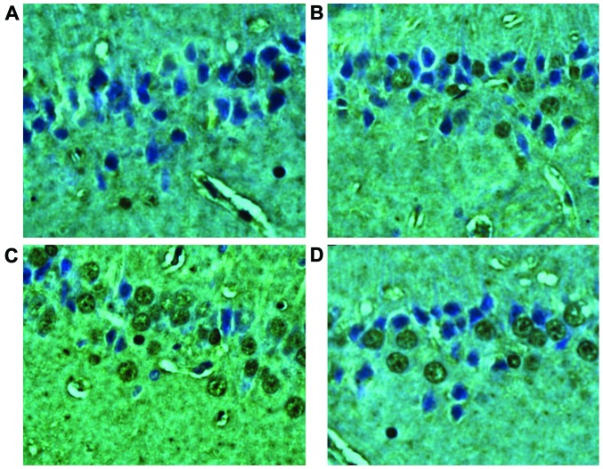
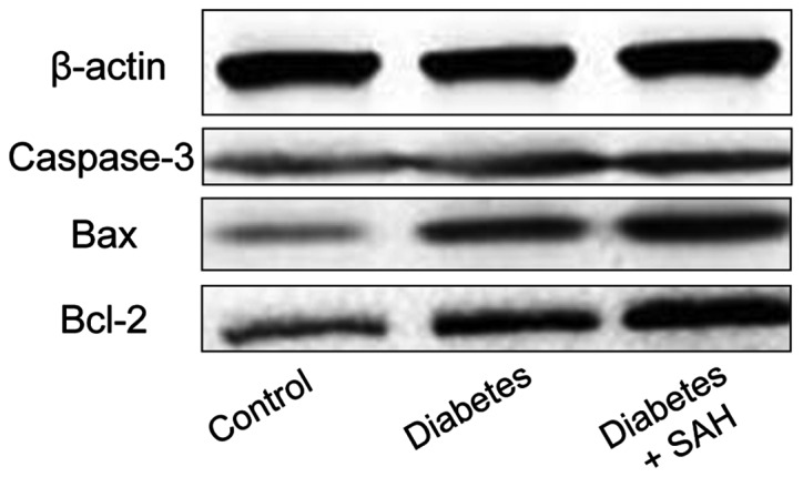
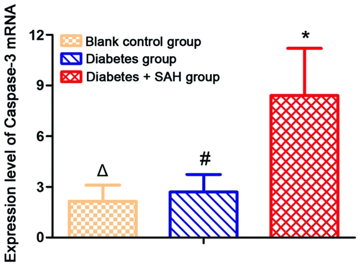
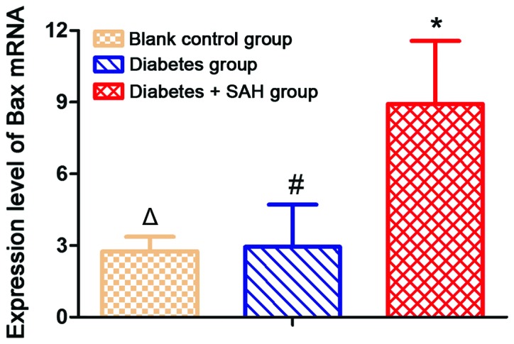
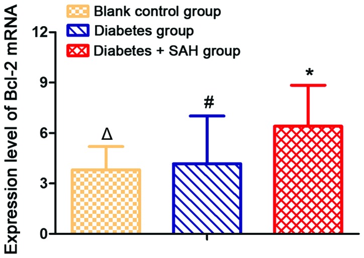
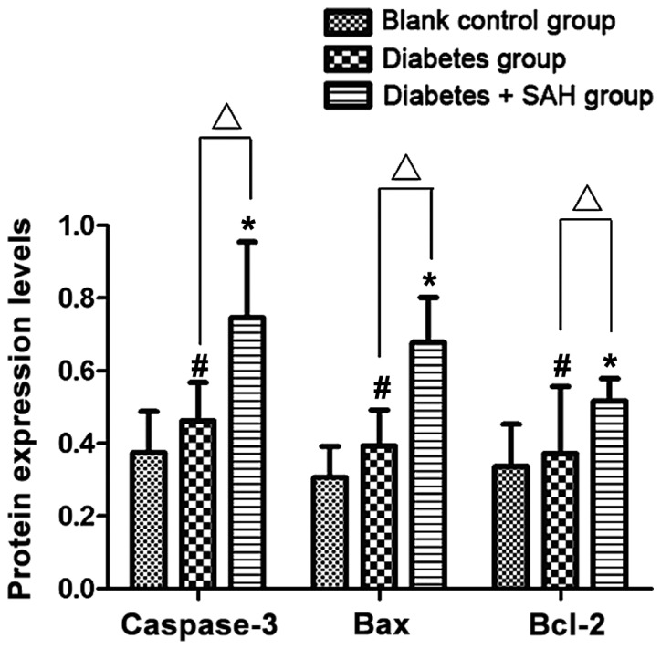
The expression of caspase-3, Bax and Bcl-2 in hippocampus of rats with diabetes and subarachnoid hemorrhage (SAH) were investigated. Diabetes mellitus model was established by intraperitoneal injection of STZ. On the basis of diabetes mellitus model, SAH animal model was established by injecting fresh autologous femoral artery blood into cerebellomedullary cisten. Rats were divided into blank control group, diabetes control group and diabetes + SAH group. TUNEL method was used to detect cell apoptosis of hippocampus. Expression levels of caspase-3, Bax and Bcl-2 were detected by real-time quantitative reverse transcription PCR and western blot analysis at mRNA and protein levels, respectively. Apoptotic cells were not detected in blank control group and diabetes group, and number of apoptotic cells was the highest in the diabetic SAH group. Expression levels of caspase-3, Bax and Bcl-2 mRNA and protein were significantly higher in diabetes + SAH group than in blank control group and diabetes group. In conclusion, Hippocampal neuron apoptosis was induced by diabetes + SAH and expression levels of caspase-3, Bax and Bcl-2 were also increased. Our study provided experimental basis for further studies of the relationship between SAH and cell apoptosis.
研究了糖尿病合并蛛网膜下腔出血(SAH)大鼠海马中半胱天冬酶-3(caspase-3)、 Bax和Bcl-2的表达。通过腹腔注射链脲佐菌素(STZ)建立糖尿病模型。在糖尿病模型的基础上,通过向小脑延髓池注射新鲜自体股动脉血建立SAH动物模型。将大鼠分为空白对照组、糖尿病对照组和糖尿病+SAH组。采用TUNEL法检测海马细胞凋亡。分别通过实时定量逆转录PCR和蛋白质免疫印迹分析在mRNA和蛋白质水平检测caspase-3、Bax和Bcl-2的表达水平。空白对照组和糖尿病组未检测到凋亡细胞,糖尿病SAH组凋亡细胞数量最多。糖尿病+SAH组中caspase-3、Bax和Bcl-2 mRNA及蛋白质的表达水平显著高于空白对照组和糖尿病组。总之,糖尿病+SAH可诱导海马神经元凋亡,且caspase-3、Bax和Bcl-2的表达水平也升高。本研究为进一步研究SAH与细胞凋亡之间的关系提供了实验依据。