Fischli Stefan, Suter-Widmer Isabelle, Nguyen Ba Tung, Müller Werner, Metzger Jürg, Strobel Klaus, Grünig Hannes, Henzen Christoph
Division of Endocrinology, Diabetes and Clinical Nutrition, Luzerner Kantonsspital, Luzern, Switzerland.
Division of Otorhinolaryngology, Head and Neck Surgery, Luzerner Kantonsspital, Luzern, Switzerland.
Front Endocrinol (Lausanne). 2018 Jan 22;8:380. doi: 10.3389/fendo.2017.00380. eCollection 2017.
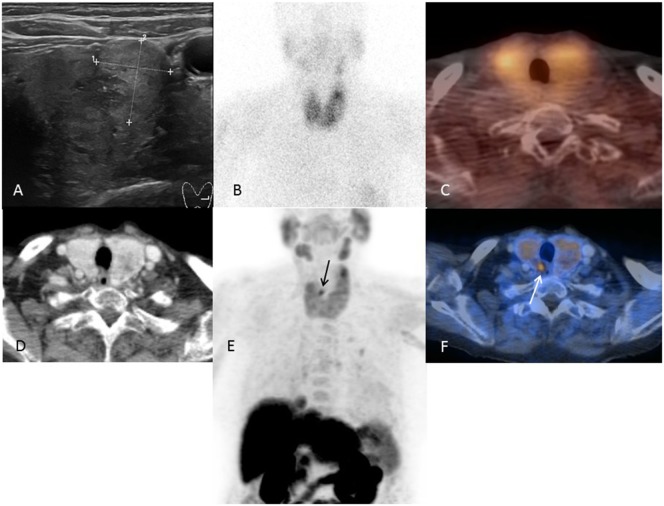
The essential prerequisite for focused parathyroidectomy in patients with primary hyperparathyroidism (pHPT) is proper localization of all autonomic tissue. Sensitivity of conventional imaging modalities (ultrasound, Tc-sestamibi scintigraphy/SPECT/CT) is influenced by different factors (i.e., size/weight and position of autonomic tissue) and decreases in the presence of a multinodular goiter. Therefore, a considerable percentage of pHPT patients have negative or equivocal localization studies before surgery. The aim of this study is to evaluate the utility of FCH-PET/CT for preoperative localization in patients with pHPT and negative/equivocal Tc-sestamibi scintigraphy/SPECT/CT and/or ultrasound.
Between 2014 and 2017, a total of 39 patients with pHPT and negative/equivocal conventional imaging were referred for FCH-PET/CT. In the analysis, we included those ( = 23) who had surgery and a histopathologic workup of the lesions.
19 of 23 patients demonstrated no tracer uptake with Tc-sestamibi scintigraphy/SPECT/CT, 6 patients had an equivocal sonographic lesion, and multinodular goiter was present in 43% (10/23). In 21 of 23 patients, hyperfunctioning parathyroid tissue was identified correctly by FCH-PET/CT [21 true positive, 1 false negative, and 1 false positive; per-patient sensitivity 95.5% (95% confidence interval {CI}, 77.2-99.9)]. 29 lesions were resected [21 true positives, 3 false negatives, 1 false positive, and 4 true negatives; per-lesion sensitivity 87.5% (95% CI, 67.6-97.3)]. All patients were classified as having surgical success according to a decrease of intraoperative parathyroid hormone of ≥50% and normalization of postoperative serum calcium levels.
Despite a high prevalence of multinodular goiter, diagnostic accuracy of FCH-PET/CT in our patient group was excellent. Therefore, FCH-PET/CT is a promising new imaging tool in patients with pHPT and negative/equivocal results by conventional imaging techniques.
对原发性甲状旁腺功能亢进症(pHPT)患者进行聚焦甲状旁腺切除术的基本前提是准确定位所有自主性甲状旁腺组织。传统成像方式(超声、锝-司他比锝显像/SPECT/CT)的敏感性受不同因素影响(如自主性甲状旁腺组织的大小/重量和位置),且在存在多结节性甲状腺肿时会降低。因此,相当一部分pHPT患者在手术前的定位检查结果为阴性或不明确。本研究的目的是评估FCH-PET/CT在术前定位pHPT且锝-司他比锝显像/SPECT/CT和/或超声检查结果为阴性/不明确的患者中的应用价值。
2014年至2017年期间,共有39例pHPT且传统成像检查结果为阴性/不明确的患者接受了FCH-PET/CT检查。在分析中,我们纳入了那些(n = 23)接受了手术及病变组织病理学检查的患者。
23例患者中,19例锝-司他比锝显像/SPECT/CT未显示示踪剂摄取,6例患者有不明确的超声病变,43%(10/23)的患者存在多结节性甲状腺肿。23例患者中有21例通过FCH-PET/CT正确识别出功能亢进的甲状旁腺组织[21例假阳性,1例假阴性,1例假阳性;患者敏感性为95.5%(95%置信区间{CI},77.2 - 99.9)]。切除了29个病变组织[21例假阳性,3例假阴性,1例假阳性,4例假阴性;病变敏感性为87.5%(95%CI,67.6 - 97.3)]。根据术中甲状旁腺激素下降≥50%及术后血清钙水平正常化,所有患者均被判定手术成功。
尽管多结节性甲状腺肿的患病率较高,但FCH-PET/CT在我们的患者组中的诊断准确性极佳。因此,对于pHPT且传统成像技术检查结果为阴性/不明确的患者,FCH-PET/CT是一种有前景的新型成像工具。