Puddighinu Giovanni, D'Amario Domenico, Foglio Eleonora, Manchi Melissa, Siracusano Andrea, Pontemezzo Elena, Cordella Martina, Facchiano Francesco, Pellegrini Laura, Mangoni Antonella, Tafani Marco, Crea Filippo, Germani Antonia, Russo Matteo Antonio, Limana Federica
Department of Experimental Medicine, Sapienza University of Rome, Rome, Italy.
Department of Cardiovascular Sciences, Catholic University of The Sacred Heart, Rome, Italy.
Oncotarget. 2017 Dec 6;9(1):937-957. doi: 10.18632/oncotarget.22946. eCollection 2018 Jan 2.
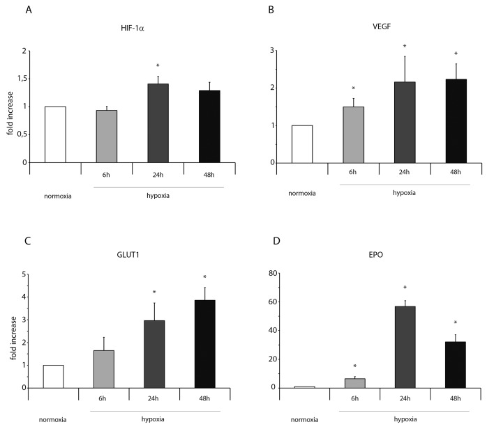
The regenerative effects of cardiac ckit stem cells (ckitCSCs) in acute myocardial infarction (MI) have been studied extensively, but how these cells exert a protective effect on cardiomyocytes is not well known. Growing evidences suggest that in adult stem cells injury triggers inflammatory signaling pathways which control tissue repair and regeneration. Aim of the present study was to determine the mechanisms underlying the cardioprotective effects of ckitCSCs following transplantation in a murine model of MI. Following isolation and expansion, cardiac ckitCSCs were subjected to normoxic and hypoxic conditions and assessed at different time points. These cells adapted to hypoxia as showed by the activation of HIF-1α and the expression of a number of genes, such as VEGF, GLUT1, EPO, HKII and, importantly, of alarmin receptors, such as RAGE, P2X7R, TLR2 and TLR4. Activation of these receptors determined an NFkB-dependent inflammatory and reparative gene response (IRR). Importantly, hypoxic ckitCSCs increased the secretion of the survival growth factors IGF-1 and HGF. To verify whether activation of the IRR in a hypoxic microenvironment could exert a beneficial effect , autologous ckitCSCs were transplanted into mouse heart following MI. Interestingly, transplantation of ckitCSCs lowered apoptotic rates and induced autophagy in the peri-infarct area; further, it reduced hypertrophy and fibrosis and, most importantly, improved cardiac function. ckitCSCs are able to adapt to a hypoxic environment and activate an inflammatory and reparative response that could account, at least in part, for a protective effect on stressed cardiomyocytes following transplantation in the infarcted heart.
心脏c-kit干细胞(ckitCSCs)在急性心肌梗死(MI)中的再生作用已得到广泛研究,但这些细胞如何对心肌细胞发挥保护作用尚不清楚。越来越多的证据表明,在成体干细胞中,损伤会触发控制组织修复和再生的炎症信号通路。本研究的目的是确定在MI小鼠模型中移植后ckitCSCs心脏保护作用的潜在机制。分离和扩增后,将心脏ckitCSCs置于常氧和低氧条件下,并在不同时间点进行评估。这些细胞适应低氧,表现为HIF-1α的激活以及许多基因的表达,如VEGF、GLUT1、EPO、HKII,重要的是,还包括警报素受体,如RAGE、P2X7R、TLR2和TLR4。这些受体的激活决定了一种依赖NFkB的炎症和修复基因反应(IRR)。重要的是,低氧ckitCSCs增加了存活生长因子IGF-1和HGF的分泌。为了验证在低氧微环境中IRR的激活是否能发挥有益作用,将自体ckitCSCs在MI后移植到小鼠心脏中。有趣的是,ckitCSCs的移植降低了梗死周边区域的凋亡率并诱导了自噬;此外,它减少了肥大和纤维化,最重要的是,改善了心脏功能。ckitCSCs能够适应低氧环境并激活炎症和修复反应,这至少可以部分解释在梗死心脏中移植后对应激心肌细胞的保护作用。