Garikipati Venkata Naga Srikanth, Singh Saurabh Pratap, Mohanram Yamuna, Gupta Ashwani Kumar, Kapoor Deepa, Nityanand Soniya
Stem Cell Research Facility, Department of Hematology, Sanjay Gandhi Post-Graduate Institute of Medical Sciences, Lucknow, India.
General Hospital, Sanjay Gandhi Post-Graduate Institute of Medical Sciences, Lucknow, India.
PLoS One. 2018 Feb 8;13(2):e0192244. doi: 10.1371/journal.pone.0192244. eCollection 2018.
Mesenchymal stem cells (MSCs) are promising cells for cardiovascular regenerative medicine. However, their potential may be limited, because of their restricted cardiovascular differentiation potential and decline in their number and functional characteristics with increasing donor age. We have previously shown that rat fetus heart harbors primitive MSCs and administration of these cells improved left ventricular (LV) function after ischemia/reperfusion injury in rats. To evaluate their potential as a new cell type for clinical cardiovascular cell therapy, we have undertaken this study on the isolation and characterization of human fetal cardiac MSCs (hfC-MSCs).
MSCs were isolated from the heart of five 14-16-week-old aborted human fetuses and studied for their growth characteristics, karyotypic stability and senescence over successive passages, expression of mesenchymal and embryonal markers by flow cytometry and immunocytochemistry, constitutive expression of cardiovascular genes by RT-PCR, differentiation into cells of the cardiovascular lineage and their immunomodulatory properties.
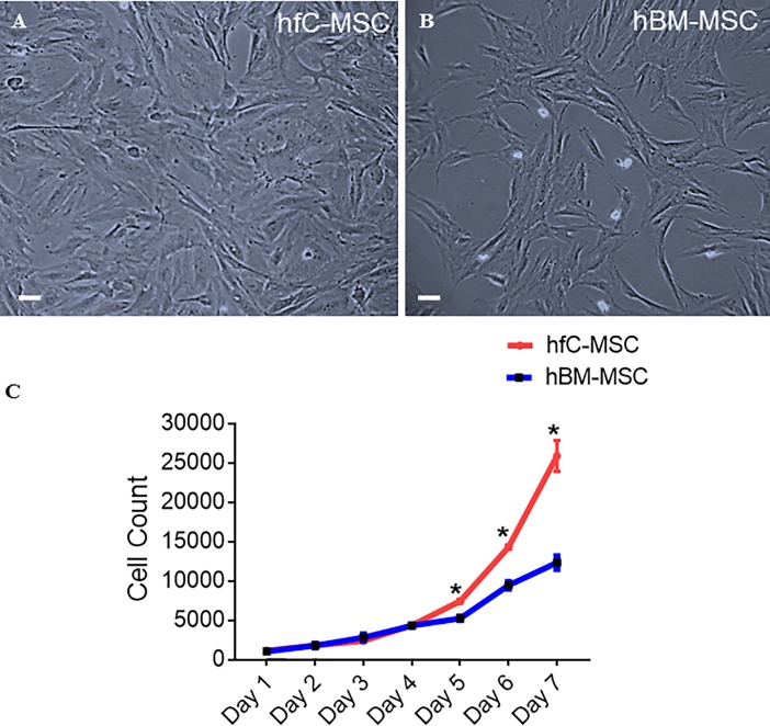
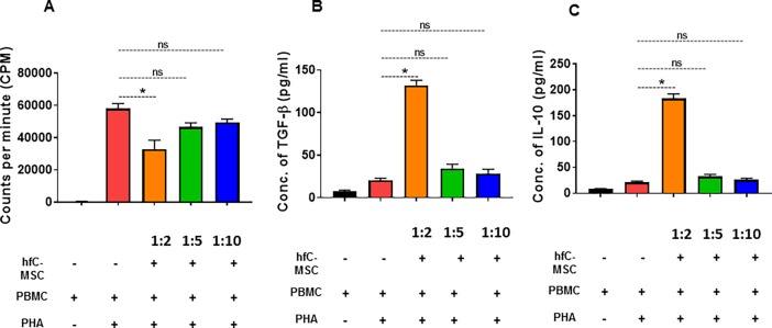
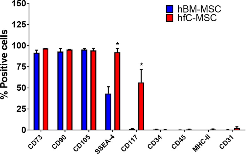
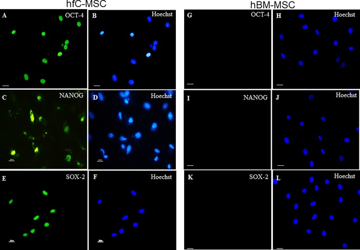
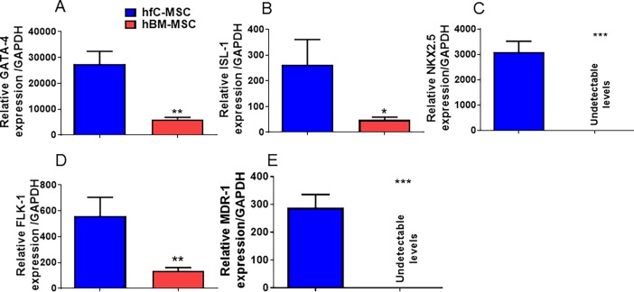
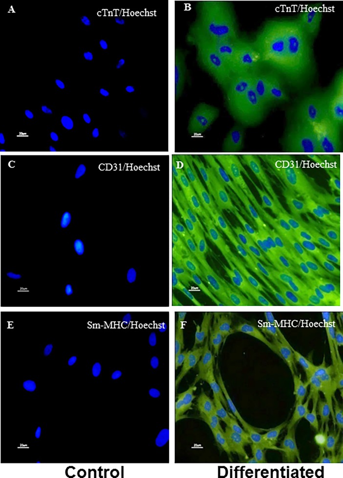
The hfC-MSCs grew as adherent monolayer with spindle shaped morphology and exhibited rapid proliferation with an average population doubling time of 34 hours and expansion to up to more than 80 population doublings with maintenance of a normal karyotype and without senescence. Immunophenotyping showed that they had similar phenotype as human bone marrow mesenchymal stem cells (hBM-MSCs) expressing CD73, CD90, CD105 and lacking expression of CD31, CD34, CD45, HLA-DR. However, hfC-MSCs expressed significantly higher levels of CD117 and SSEA-4 compared to hBM-MSCs. In addition, hfC-MSCs expressed the embryonal markers Oct-4, Nanog and Sox-2 as compared to hBM-MSCs. Further, hfC-MSCs had significantly higher expression of the cardiovascular genes viz. ISL-1, flk-1, GATA-4, NKX2.5 and MDR-1 as compared to hBM-MSCs, and could be differentiated into major cardiovascular cells (cardiomyocytes, endothelial cells, smooth muscle cells). Interestingly, hfC-MSCs markedly reduced T-lymphocyte proliferation with an increased secretion of TGF-β and IL-10.
Our results show that human fetus heart is a novel source of primitive MSCs with cardiovascular commitment which may have a potential therapeutic application in cardiovascular regenerative medicine.
间充质干细胞(MSCs)是心血管再生医学中很有前景的细胞。然而,由于其有限的心血管分化潜能以及随着供体年龄增长其数量和功能特性的下降,它们的潜力可能受到限制。我们之前已经表明,大鼠胎儿心脏含有原始间充质干细胞,给予这些细胞可改善大鼠缺血/再灌注损伤后的左心室(LV)功能。为了评估它们作为临床心血管细胞治疗新细胞类型的潜力,我们开展了这项关于人胎儿心脏间充质干细胞(hfC-MSCs)分离和特性分析的研究。
从5例14 - 16周大人工流产胎儿的心脏中分离间充质干细胞,并研究其生长特性、连续传代过程中的核型稳定性和衰老情况,通过流式细胞术和免疫细胞化学检测间充质和胚胎标志物的表达,通过逆转录聚合酶链反应(RT-PCR)检测心血管基因的组成性表达,向心血管谱系细胞的分化及其免疫调节特性。
hfC-MSCs以贴壁单层生长,呈纺锤形形态,增殖迅速,平均群体倍增时间为34小时,可扩增至80多个群体倍增,维持正常核型且无衰老。免疫表型分析显示,它们与人类骨髓间充质干细胞(hBM-MSCs)具有相似的表型,表达CD73、CD90、CD105,不表达CD31、CD34、CD45、HLA-DR。然而,与hBM-MSCs相比,hfC-MSCs表达的CD117和SSEA-4水平显著更高。此外,与hBM-MSCs相比,hfC-MSCs表达胚胎标志物Oct-4、Nanog和Sox-2。进一步地,与hBM-MSCs相比,hfC-MSCs的心血管基因即ISL-1、flk-1、GATA-4、NKX2.5和MDR-1的表达显著更高,并且可以分化为主要的心血管细胞(心肌细胞、内皮细胞、平滑肌细胞)。有趣的是,hfC-MSCs显著降低T淋巴细胞增殖,同时TGF-β和IL-10的分泌增加。
我们的结果表明,人胎儿心脏是具有心血管定向分化能力的原始间充质干细胞的新来源,可能在心血管再生医学中具有潜在的治疗应用价值。