Department of Neurosurgery, Emory University School of Medicine, Atlanta, GA.
Department of Neurology, Emory University School of Medicine, Atlanta, GA.
Ann Neurol. 2018 Mar;83(3):575-587. doi: 10.1002/ana.25180. Epub 2018 Mar 3.
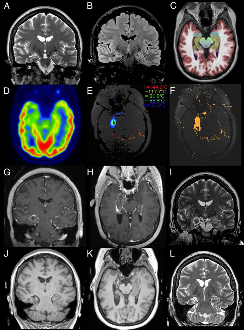
To evaluate the outcomes 1 year and longer following stereotactic laser amygdalohippocampotomy for mesial temporal lobe epilepsy in a large series of patients treated over a 5-year period since introduction of this novel technique.
Surgical outcomes of a consecutive series of 58 patients with mesial temporal lobe epilepsy who underwent the surgery at our institution with at least 12 months of follow-up were retrospectively evaluated. A subgroup analysis was performed comparing patients with and without mesial temporal sclerosis.
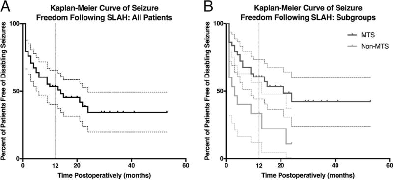
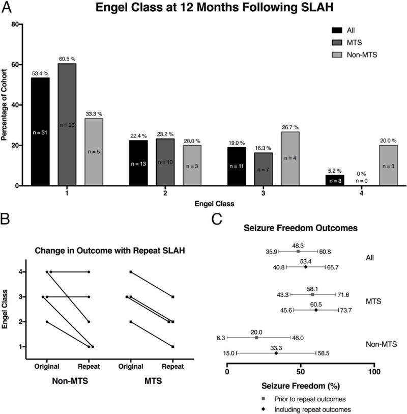
One year following stereotactic laser amygdalohippocampotomy, 53.4% (95% confidence interval [CI] = 40.8-65.7%) of all patients were free of disabling seizures (Engel I). Three of 9 patients became seizure-free following repeat ablation. Subgroup analysis showed that 60.5% (95% CI = 45.6-73.7%) of patients with mesial temporal sclerosis were free of disabling seizures as compared to 33.3% (95% CI = 15.0-58.5%) of patients without mesial temporal sclerosis. Quality of Life in Epilepsy-31 scores significantly improved at the group level, few procedure-related complications were observed, and verbal memory outcome was better than historical open resection data.
In an unselected consecutive series of patients, stereotactic laser amygdalohippocampotomy yielded seizure-free rates for patients with mesial temporal lobe epilepsy lower than, but comparable to, the outcomes typically associated with open temporal lobe surgery. Analogous to results from open surgery, patients without mesial temporal sclerosis fared less well. This novel procedure is an effective minimally invasive alternative to resective surgery. In the minority of patients not free of disabling seizures, laser ablation presents no barrier to additional open surgery. Ann Neurol 2018;83:575-587.
评估在立体定向激光杏仁核海马切开术引入后的 5 年期间,对一系列接受该新技术治疗的患者进行长达 1 年以上的随访后,其治疗结果。
回顾性分析了在我院接受手术且随访时间至少 12 个月的 58 例内侧颞叶癫痫患者的连续系列手术结果。对有或无内侧颞叶硬化的患者进行了亚组分析。
立体定向激光杏仁核海马切开术后 1 年,所有患者中有 53.4%(95%置信区间[CI] = 40.8-65.7%)无残疾性发作(Engel I 级)。9 例中有 3 例在重复消融后无发作。亚组分析显示,内侧颞叶硬化患者中有 60.5%(95%CI = 45.6-73.7%)无残疾性发作,而无内侧颞叶硬化患者中有 33.3%(95%CI = 15.0-58.5%)。癫痫生活质量 31 项评分在组内显著提高,仅观察到少数与手术相关的并发症,且言语记忆结果优于历史上的开放性切除术数据。
在一项未选择的连续患者系列中,立体定向激光杏仁核海马切开术治疗内侧颞叶癫痫的无发作率低于,但与开放式颞叶手术相关的结果相当。与开放式手术结果类似,无内侧颞叶硬化的患者效果较差。这种新的手术方法是一种有效的微创替代切除手术。在大多数无残疾性发作的患者中,激光消融不会对额外的开放性手术造成障碍。