Han Hailin, Wang Dongmei, Yang Maowu, Wang Shenhao
Department of Radiology, The Second People's Hospital of Liaocheng Affiliated to Taishan Medical College, Liaocheng, Shandong 252600, P.R. China.
Department of Gastroenterology, The Second People's Hospital of Liaocheng Affiliated to Taishan Medical College, Liaocheng, Shandong 252600, P.R. China.
Oncol Lett. 2018 Feb;15(2):2073-2078. doi: 10.3892/ol.2017.7539. Epub 2017 Dec 6.
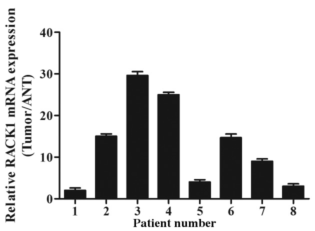
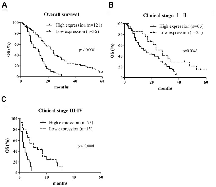
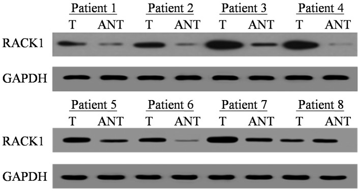
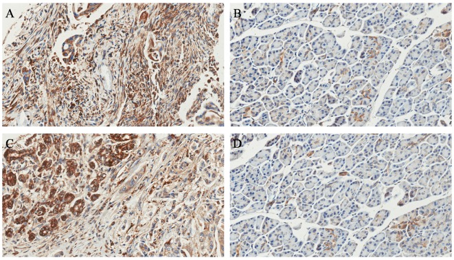
Receptor for activated C kinase 1 (RACK1) is associated with certain aspects of cancer biology and signaling pathways, but its function in pancreatic ductal adenocarcinoma (PDAC) remains unknown. In the present study, 157 patients with PDAC were enrolled. RACK1 mRNA and protein expression levels were analyzed in PDAC tissues and matched adjacent noncancerous tissues by reverse transcription-quantitative polymerase chain reaction and western blotting. RACK1 expression levels in paraffin-embedded PDAC tissues were determined by immunohistochemistry. The associations between RACK1 expression and clinical data were evaluated using χ analysis. The relationship between RACK1 expression and the survival data of patients was analyzed using Kaplan-Meier and log rank tests. RACK1 mRNA and protein were revealed to be overexpressed in PDAC tumor tissues compared with adjacent noncancerous tissues. RACK1 expression was associated with clinical stage (P=0.001), lymph node invasion (P=0.003) and liver metastasis (P=0.001). Furthermore, patients with PDAC and high RACK1 expression demonstrated shorter overall survival times compared with patients with low RACK1 expression (P=0.002). Multivariate analysis indicated that RACK1 overexpression was an independent prognostic factor for patients with PDAC. Overexpression of RACK1 may contribute to tumor progression, and may be a potential prognostic biomarker for patients with PDAC.
活化C激酶1受体(RACK1)与癌症生物学和信号通路的某些方面相关,但其在胰腺导管腺癌(PDAC)中的功能尚不清楚。在本研究中,纳入了157例PDAC患者。通过逆转录定量聚合酶链反应和蛋白质印迹法分析了PDAC组织及配对的癌旁非癌组织中RACK1 mRNA和蛋白表达水平。采用免疫组织化学法测定石蜡包埋的PDAC组织中RACK1表达水平。使用χ分析评估RACK1表达与临床数据之间的关联。采用Kaplan-Meier法和对数秩检验分析RACK1表达与患者生存数据之间的关系。结果显示,与癌旁非癌组织相比,RACK1 mRNA和蛋白在PDAC肿瘤组织中过表达。RACK1表达与临床分期(P = 0.001)、淋巴结浸润(P = 0.003)和肝转移(P = 0.001)相关。此外,与RACK1低表达患者相比,RACK1高表达的PDAC患者总生存时间更短(P = 0.002)。多因素分析表明,RACK1过表达是PDAC患者的独立预后因素。RACK1过表达可能促进肿瘤进展,可能是PDAC患者潜在的预后生物标志物。