Lundberg Marcus, Stenwall Anton, Tegehall Angie, Korsgren Olle, Skog Oskar
a Department of Immunology, Genetics and Pathology , Uppsala University , Uppsala , Sweden.
b Department of Clinical Chemistry and Transfusion Medicine , Institute of Biomedicine, University of Gothenburg , Gothenburg , Sweden.
Islets. 2018 Mar 4;10(2):69-79. doi: 10.1080/19382014.2018.1433980. Epub 2018 Feb 15.
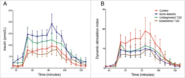
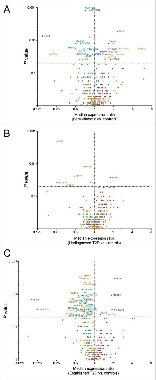
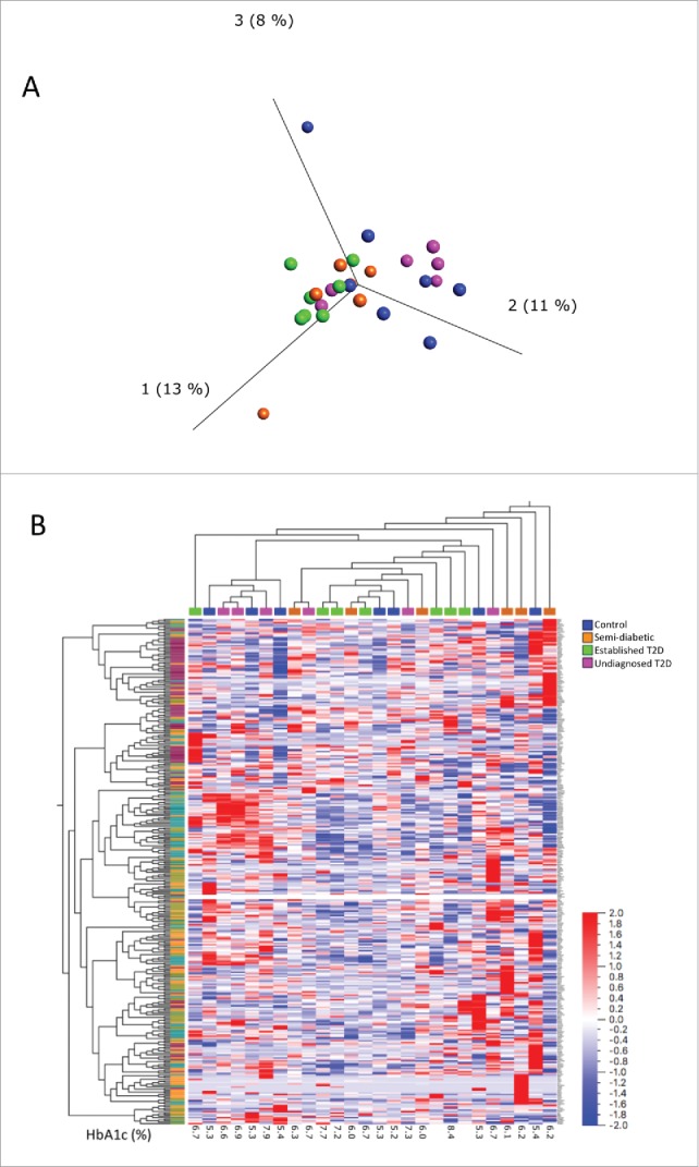
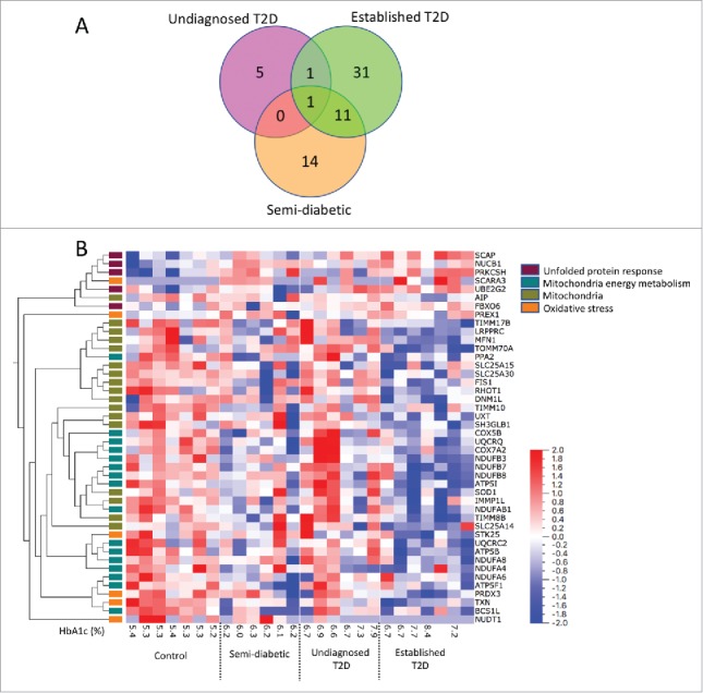
It is currently unknown how the islet transcriptional pattern changes as glucose metabolism deteriorates and progresses to fulminant type 2 diabetes (T2D). In this study, we hypothesized that islets from donors with elevated HbA1c levels, but not yet diagnosed with T2D, would show signs of cell stress on a transcriptional level. Laser capture microdissection and qPCR arrays including 330 genes related to mitochondria, oxidative stress, or the unfolded protein response were used to extract and analyze islets from organ donors with HbA1c <5.5% (37 mmol/mol), elevated HbA1c (6.0-6.5% (42-48 mmol/mol)), high HbA1c (>6.5% (48 mmol/mol)) or established T2D. Principal component analysis and hierarchical clustering based on the expression of all 330 genes displayed no obvious separation of the four different donor groups, indicating that the inter-donor variations were larger than the differences between groups. However, 44 genes were differentially expressed (P < 0.05, false discovery rate <30%) between islets from donors with HbA1c <5.5% (37 mmol/mol) compared with islets from T2D subjects. Twelve genes were differentially expressed compared to control islets in both donors with established T2D and donors with elevated HbA1c (6.0-6.5% (42-48 mmol/mol)). Overexpressed genes were related mainly to the unfolded protein response, whereas underexpressed genes were related to mitochondria. Our data on transcriptional changes in human islets retrieved by LCM from high-quality biopsies, as pre-diabetes progresses to established T2D, increase our understanding on how islet stress contributes to the disease development.
目前尚不清楚随着葡萄糖代谢恶化并进展为暴发性2型糖尿病(T2D),胰岛转录模式如何变化。在本研究中,我们假设来自糖化血红蛋白(HbA1c)水平升高但尚未诊断为T2D的供体的胰岛在转录水平上会显示细胞应激迹象。使用激光捕获显微切割和qPCR阵列(包括330个与线粒体、氧化应激或未折叠蛋白反应相关的基因)从HbA1c<5.5%(37 mmol/mol)、HbA1c升高(6.0 - 6.5%(42 - 48 mmol/mol))、高HbA1c(>6.5%(48 mmol/mol))或已确诊T2D的器官供体中提取并分析胰岛。基于所有330个基因的表达进行主成分分析和层次聚类,未显示出四个不同供体组之间有明显分离,这表明供体间差异大于组间差异。然而,与T2D受试者的胰岛相比,HbA1c<5.5%(37 mmol/mol)的供体的胰岛中有44个基因差异表达(P<0.05,错误发现率<30%)。在已确诊T2D的供体和HbA1c升高(6.0 - 6.5%(42 - 48 mmol/mol))的供体中,与对照胰岛相比,均有12个基因差异表达。过表达的基因主要与未折叠蛋白反应相关,而低表达的基因与线粒体相关。我们从高质量活检组织中通过激光捕获显微切割获得的关于人类胰岛转录变化的数据,随着糖尿病前期进展为确诊的T2D,增加了我们对胰岛应激如何促进疾病发展的理解。