Gupta Rajeev, Ghosh Subhendu
Department of Physiology, All India Institute of Medical Sciences, India.
Department of Biophysics, University of Delhi South Campus, India.
Biochim Open. 2017 Feb 11;4:41-46. doi: 10.1016/j.biopen.2017.02.001. eCollection 2017 Jun.
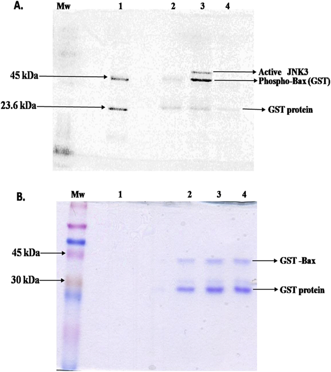
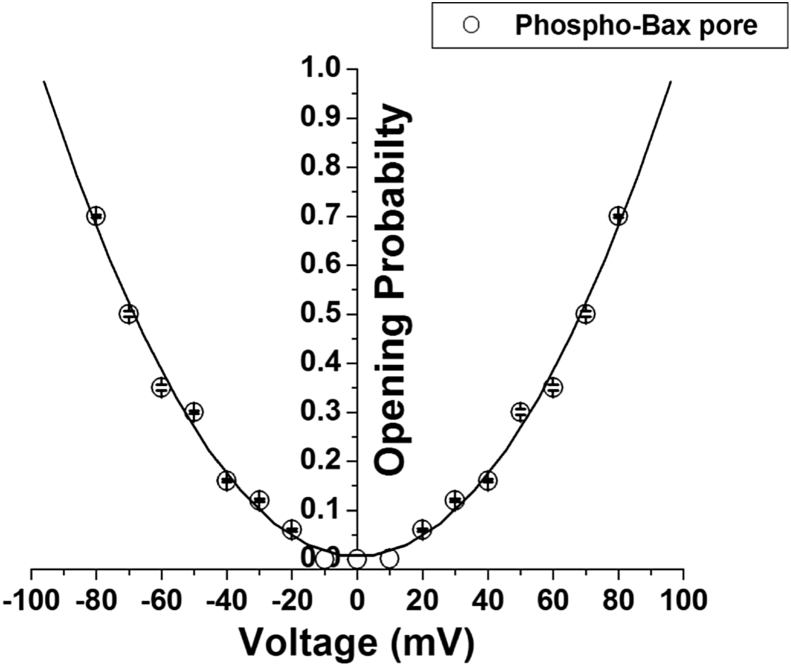
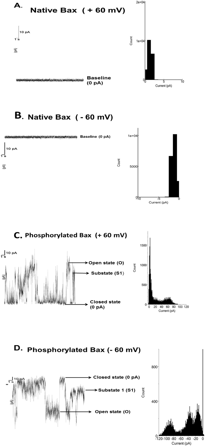
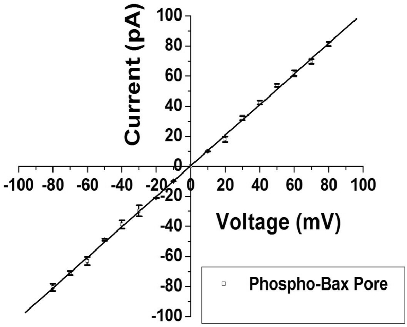
Bax is a pro-apoptotic cytosolic protein. In this work native (unphosphorylated) and JNK3 phosphorylated Bax proteins are studied on artificial bilayer membranes for pore formation. Phosphorylated Bax formed pore on the bilayer lipid membrane whereas native one does not. In cells undergoing apoptosis the pore formed by the phosphorylated Bax could be important in cytochrome release from the mitochondrial intermembrane space to the cytosol. The low conductance (1.5 nS) of the open state of the phosphorylated Bax pore corresponds to pore diameter of 0.9 nm which is small to release cytochrome (∼3.4 nm). We hypothesized that JNK3 phosphorylated Bax protein can form bigger pores after forming complexes with other mitochondrial proteins like VDAC, t-Bid etc. to release cytochrome .
Bax是一种促凋亡的胞质蛋白。在这项研究中,对天然(未磷酸化)和JNK3磷酸化的Bax蛋白在人工双层膜上形成孔的情况进行了研究。磷酸化的Bax在双层脂质膜上形成孔,而天然的Bax则不能。在经历凋亡的细胞中,磷酸化的Bax形成的孔对于细胞色素从线粒体膜间隙释放到细胞质中可能很重要。磷酸化的Bax孔开放状态下的低电导率(1.5 nS)对应于0.9 nm的孔径,该孔径太小以至于无法释放细胞色素(约3.4 nm)。我们推测,JNK3磷酸化的Bax蛋白在与其他线粒体蛋白(如VDAC、t-Bid等)形成复合物后可以形成更大的孔,从而释放细胞色素。