Ylitalo Pekka, Pitkänen Olli M, Lauerma Kirsi, Holmström Miia, Rahkonen Otto, Heikinheimo Markku, Sairanen Heikki, Jokinen Eero
Children's Hospital, University of Helsinki and Helsinki University Central Hospital, Helsinki, Finland.
Helsinki Medical Imaging Center, Helsinki University Central Hospital, Helsinki, Finland.
Int J Cardiol Heart Vessel. 2014 Feb 12;3:15-20. doi: 10.1016/j.ijchv.2014.01.002. eCollection 2014 Jun.
Fibrosis after myocardial damage can be determined by cardiac magnetic resonance (CMR) with late gadolinium enhancement (LGE). We studied whether ventricular LGE is visible in the ventricles of pediatric and adolescent TOF (tetralogy of Fallot) patients by measuring LGE and investigating whether fibrosis correlated with right ventricular volume, pulmonary regurgitation, N-terminal pro-brain natriuretic peptide (NT-proBNP) or the aminoterminal propeptide of type III procollagen (PIIINP). We also studied if the patient's age, post-operative follow-up time or surgical history would affect LGE.
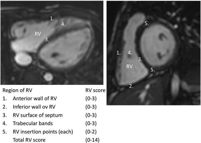
A total of 40 pediatric patients who had undergone TOF repair and 43 healthy age and gender matched controls underwent a CMR study, whereby LGE was scored in the right (RV) and the left ventricle. To exclude the possible iatrogenic scarring we calculated the LGE score by excluding the right ventricular outflow tract and VSD patch region.
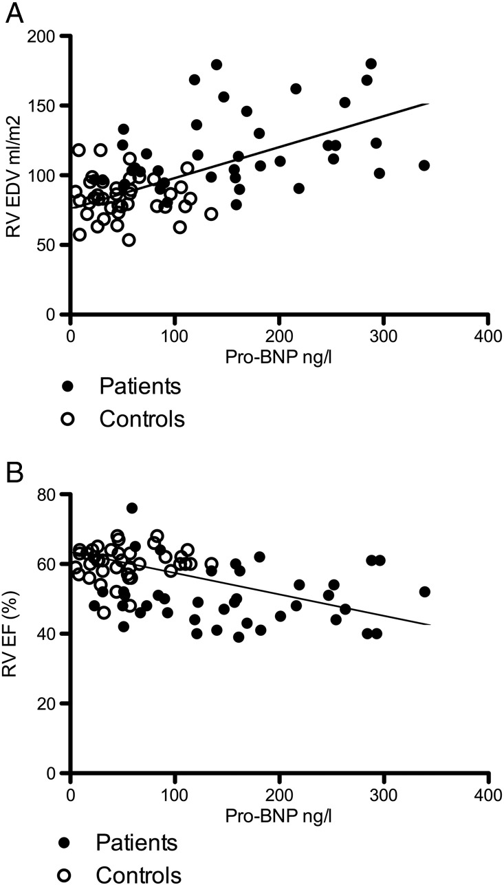
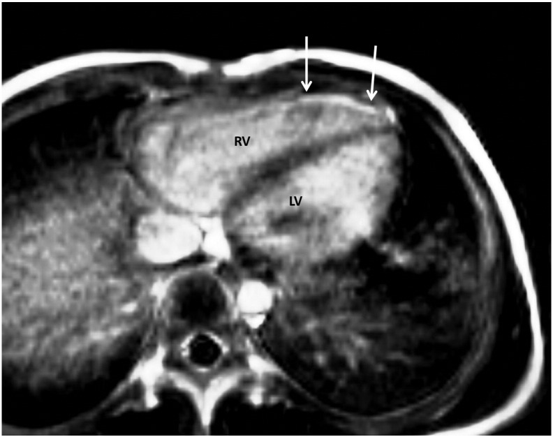
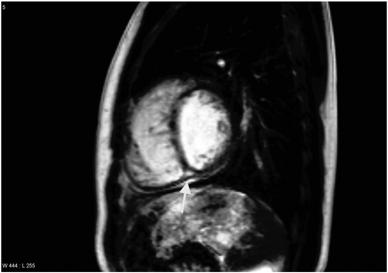
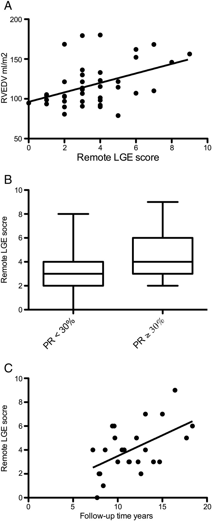
All patients had RV LGE and in 39 of 40 it was seen also outside the surgically affected areas. The amount of LGE correlated positively with the RV end-diastolic volume (r = 0.44, P = 0.0045), pulmonary regurgitation (r = 0.40, P = 0.013), and with NT-proBNP. The presence of LGE also depended on post-operative follow-up time (r = 0.53, P = 0.006). PIIINP levels of TOF patients were significantly higher than in the control subjects but it did not correlate with LGE or with any of the studied clinical markers.
LGE is present globally in the right ventricular muscle in children and adolescents with TOF. The longer the follow-up time the more common was the LGE in the right ventricle.
心肌损伤后的纤维化可通过心脏磁共振成像(CMR)及延迟钆增强(LGE)来确定。我们通过测量LGE并研究纤维化是否与右心室容积、肺动脉反流、N末端脑钠肽前体(NT-proBNP)或III型前胶原氨基端前肽(PIIINP)相关,来研究小儿及青少年法洛四联症(TOF)患者心室中是否可见心室LGE。我们还研究了患者年龄、术后随访时间或手术史是否会影响LGE。
共有40例接受TOF修复术的小儿患者和43例年龄及性别匹配的健康对照者接受了CMR检查,对右心室(RV)和左心室的LGE进行评分。为排除可能的医源性瘢痕形成,我们通过排除右心室流出道和室间隔缺损补片区域来计算LGE评分。
所有患者均有右心室LGE,40例中有39例在手术影响区域以外也可见LGE。LGE的量与右心室舒张末期容积呈正相关(r = 0.44,P = 0.0045)、与肺动脉反流呈正相关(r = 0.40,P = 0.013),并与NT-proBNP呈正相关。LGE的存在还取决于术后随访时间(r = 0.53,P = 0.006)。TOF患者的PIIINP水平显著高于对照组,但它与LGE或任何研究的临床指标均无相关性。
LGE在患有TOF的儿童和青少年的右心室心肌中普遍存在。随访时间越长,右心室中LGE越常见。