• 文献检索
  • 文档翻译
  • 深度研究
  • 学术资讯
  • Suppr Zotero 插件Zotero 插件
  • 邀请有礼
  • 套餐&价格
  • 历史记录
应用&插件
Suppr Zotero 插件Zotero 插件浏览器插件Mac 客户端Windows 客户端微信小程序
定价
高级版会员购买积分包购买API积分包
服务
文献检索文档翻译深度研究API 文档MCP 服务
关于我们
关于 Suppr公司介绍联系我们用户协议隐私条款
关注我们

Suppr 超能文献

核心技术专利:CN118964589B侵权必究
粤ICP备2023148730 号-1Suppr @ 2026

文献检索

告别复杂PubMed语法,用中文像聊天一样搜索,搜遍4000万医学文献。AI智能推荐,让科研检索更轻松。

立即免费搜索

文件翻译

保留排版,准确专业,支持PDF/Word/PPT等文件格式,支持 12+语言互译。

免费翻译文档

深度研究

AI帮你快速写综述,25分钟生成高质量综述,智能提取关键信息,辅助科研写作。

立即免费体验

扩散加权成像在识别乳腺癌对新辅助化疗的病理反应中的应用:一项荟萃分析。

Diffusion-weighted imaging in identifying breast cancer pathological response to neoadjuvant chemotherapy: A meta-analysis.

作者信息

Chu Wei, Jin Weiwei, Liu Daihong, Wang Jian, Geng Chengjun, Chen Lihua, Huang Xuequan

机构信息

Department of Radiology, Wuxi Huishan District People's Hospital, Jiangsu Province, 214187, China.

Department of Radiology, Wuxi Second Traditional Chinese Medicine Hospital, Jiangsu Province, 214121, China.

出版信息

Oncotarget. 2017 Dec 11;9(6):7088-7100. doi: 10.18632/oncotarget.23195. eCollection 2018 Jan 23.

DOI:10.18632/oncotarget.23195
PMID:29467952
原文链接:https://pmc.ncbi.nlm.nih.gov/articles/PMC5805538/
Abstract

BACKGROUND

Diffusion-weighted imaging (DWI) is increasingly used to identify pathological complete responses (pCRs) to neoadjuvant chemotherapy (NAC) in breast cancer. The aim of the present study was to assess the utility of DWI using a pooled analysis.

MATERIALS AND METHODS

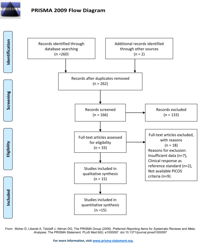
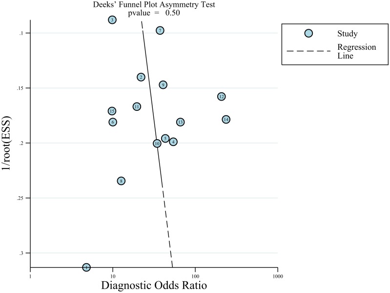
Literature databases were searched prior to July 2017. Fifteen studies with a total of 1181 patients were included. The data were extracted to perform pooled analysis, heterogeneity testing, threshold effect testing, sensitivity analysis, publication bias analysis and subgroup analyses.

RESULT

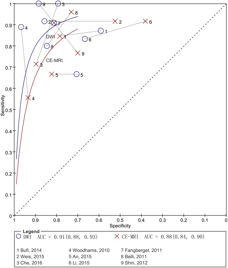
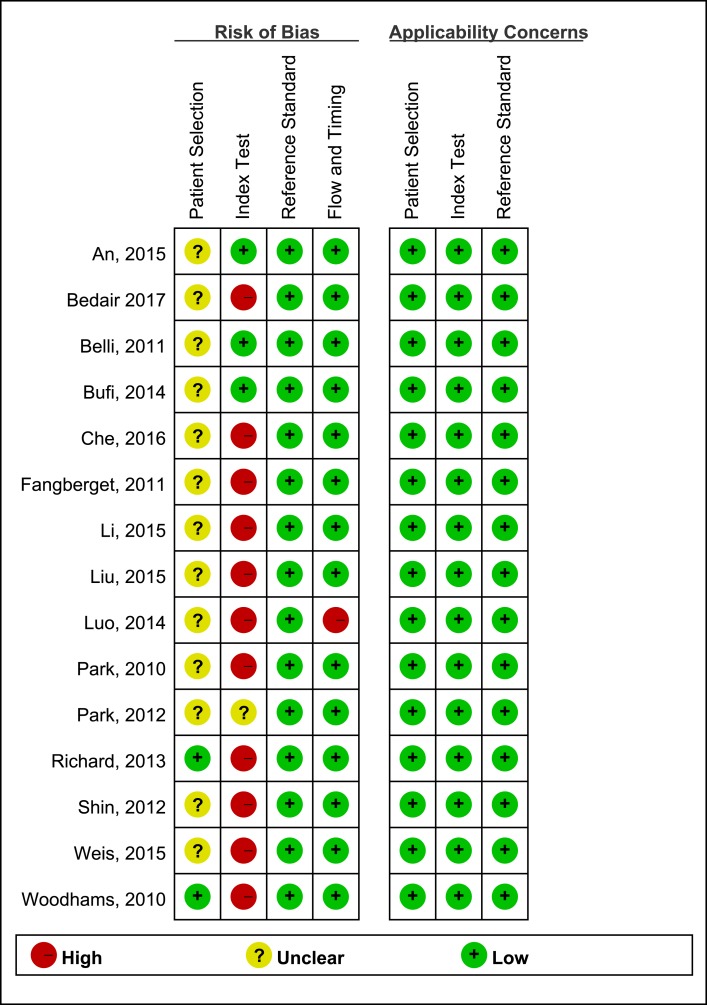
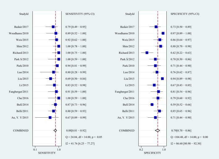
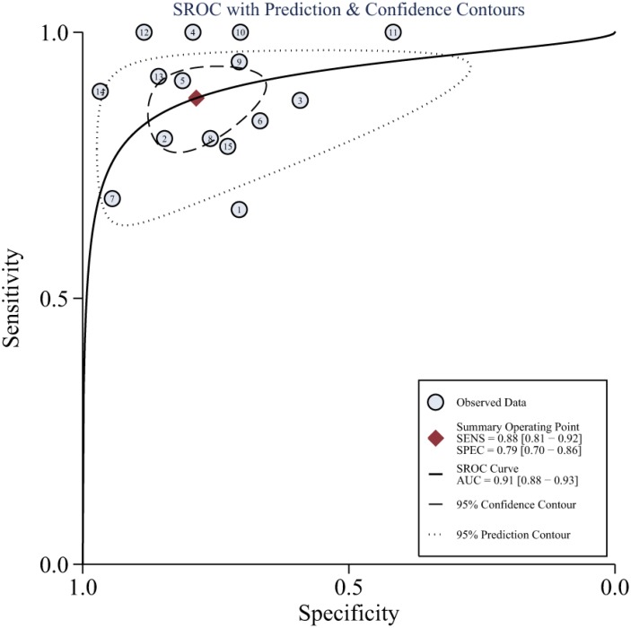
The methodological quality was moderate. Remarkable heterogeneity was detected, primarily due to a threshold effect. The pooled weighted values were a sensitivity of 0.88 (95% confidence interval (CI): 0.81, 0.92), a specificity of 0.79 (95% CI: 0.70, 0.86), a positive likelihood ratio of 4.1 (95% CI: 2.9, 5.9), a negative likelihood ratio of 0.16 (95% CI: 0.10, 0.24), and a diagnostic odds ratio of 26 (95% CI: 15, 46). The area under the receiver operator characteristic curve was 0.91 (95% CI: 0.88, 0.93). In the subgroup analysis, the pooled specificity of change in the apparent diffusion coefficient (ADC) subgroup was higher than that in the pre-treatment ADC subgroup (0.80 [95% CI: 0.71, 087] vs. 0.63 [95% CI: 0.52, 0.73], = 0.027).

CONCLUSIONS

DWI may be an accurate and nonradioactive imaging technique for identifying pCRs to NAC in breast cancer. Nonetheless, there are a variety of issues when assessing DWI techniques for estimating breast cancer responses to NAC, and large scale and well-designed clinical trials are needed to assess the technique's diagnostic value.

摘要

背景

弥散加权成像(DWI)越来越多地用于识别乳腺癌新辅助化疗(NAC)后的病理完全缓解(pCR)。本研究的目的是通过汇总分析评估DWI的效用。

材料与方法

检索2017年7月之前的文献数据库。纳入15项研究,共1181例患者。提取数据进行汇总分析、异质性检验、阈值效应检验、敏感性分析、发表偏倚分析和亚组分析。

结果

方法学质量中等。检测到显著的异质性,主要归因于阈值效应。汇总加权值为灵敏度0.88(95%置信区间(CI):0.81,0.92),特异度0.79(95%CI:0.70,0.86),阳性似然比4.1(95%CI:2.9,5.9),阴性似然比0.16(95%CI:0.10,0.24),诊断比值比26(95%CI:15,46)。受试者工作特征曲线下面积为0.91(95%CI:0.88,0.93)。在亚组分析中,表观扩散系数(ADC)变化亚组的汇总特异度高于治疗前ADC亚组(0.80[95%CI:0.71,0.87]对0.63[95%CI:0.52,0.73],P = 0.027)。

结论

DWI可能是一种准确且无辐射的成像技术,用于识别乳腺癌对NAC的pCR。尽管如此,在评估DWI技术以估计乳腺癌对NAC的反应时存在各种问题,需要大规模且设计良好的临床试验来评估该技术的诊断价值。

https://cdn.ncbi.nlm.nih.gov/pmc/blobs/b0a1/5805538/a5b3dce4ec3f/oncotarget-09-7088-g006.jpg
https://cdn.ncbi.nlm.nih.gov/pmc/blobs/b0a1/5805538/c6970ee5abbf/oncotarget-09-7088-g001.jpg
https://cdn.ncbi.nlm.nih.gov/pmc/blobs/b0a1/5805538/d8384f1ca263/oncotarget-09-7088-g002.jpg
https://cdn.ncbi.nlm.nih.gov/pmc/blobs/b0a1/5805538/20e24ab8d404/oncotarget-09-7088-g003.jpg
https://cdn.ncbi.nlm.nih.gov/pmc/blobs/b0a1/5805538/5799aa0a8c4a/oncotarget-09-7088-g004.jpg
https://cdn.ncbi.nlm.nih.gov/pmc/blobs/b0a1/5805538/5bac60eadd21/oncotarget-09-7088-g005.jpg
https://cdn.ncbi.nlm.nih.gov/pmc/blobs/b0a1/5805538/a5b3dce4ec3f/oncotarget-09-7088-g006.jpg

相似文献

1
Diffusion-weighted imaging in identifying breast cancer pathological response to neoadjuvant chemotherapy: A meta-analysis.扩散加权成像在识别乳腺癌对新辅助化疗的病理反应中的应用:一项荟萃分析。
Oncotarget. 2017 Dec 11;9(6):7088-7100. doi: 10.18632/oncotarget.23195. eCollection 2018 Jan 23.
2
Diffusion-weighted imaging in monitoring the pathological response to neoadjuvant chemotherapy in patients with breast cancer: a meta-analysis.弥散加权成像监测乳腺癌新辅助化疗病理反应的meta 分析。
World J Surg Oncol. 2018 Jul 18;16(1):145. doi: 10.1186/s12957-018-1438-y.
3
A systematic review and meta-analysis of the accuracy of diffusion-weighted MRI in the detection of malignant pulmonary nodules and masses.扩散加权 MRI 检测恶性肺结节和肿块的准确性的系统评价和荟萃分析。
Acad Radiol. 2014 Jan;21(1):21-9. doi: 10.1016/j.acra.2013.09.019.
4
Diffusion-weighted imaging in assessing pathological response of tumor in breast cancer subtype to neoadjuvant chemotherapy.扩散加权成像在评估乳腺癌亚型肿瘤对新辅助化疗的病理反应中的应用
J Magn Reson Imaging. 2015 Sep;42(3):779-87. doi: 10.1002/jmri.24843. Epub 2015 Jan 8.
5
The diagnostic performance of diffusion-weighted imaging and dynamic contrast-enhanced magnetic resonance imaging in evaluating the pathological response of breast cancer to neoadjuvant chemotherapy: A meta-analysis.扩散加权成像和动态对比增强磁共振成像在评估乳腺癌新辅助化疗病理反应中的诊断性能:荟萃分析。
Eur J Radiol. 2021 Oct;143:109931. doi: 10.1016/j.ejrad.2021.109931. Epub 2021 Aug 23.
6
Meta-analysis of diffusion-weighted MRI in the differential diagnosis of lung lesions.弥散加权 MRI 对肺部病变的鉴别诊断的荟萃分析。
J Magn Reson Imaging. 2013 Jun;37(6):1351-8. doi: 10.1002/jmri.23939. Epub 2012 Dec 12.
7
Diffusion-weighted MRI in differentiating malignant from benign thyroid nodules: a meta-analysis.磁共振扩散加权成像在鉴别甲状腺良恶性结节中的应用:一项荟萃分析
BMJ Open. 2016 Jan 5;6(1):e008413. doi: 10.1136/bmjopen-2015-008413.
8
Evaluation of the efficacy of neoadjuvant chemotherapy for breast cancer using diffusion-weighted imaging and dynamic contrast-enhanced magnetic resonance imaging.使用扩散加权成像和动态对比增强磁共振成像评估乳腺癌新辅助化疗的疗效。
Neoplasma. 2017;64(3):430-436. doi: 10.4149/neo_2017_314.
9
Diagnostic value of whole-body diffusion-weighted magnetic resonance imaging for detection of primary and metastatic malignancies: a meta-analysis.全身扩散加权磁共振成像对原发性和转移性恶性肿瘤检测的诊断价值:一项荟萃分析。
Eur J Radiol. 2014 Feb;83(2):338-44. doi: 10.1016/j.ejrad.2013.11.017. Epub 2013 Dec 4.
10
Meta-Analysis of MRI in Predicting Early Response to Radiotherapy and Chemotherapy in Esophageal Cancer.MRI预测食管癌放疗和化疗早期反应的Meta分析
Acad Radiol. 2025 Feb;32(2):798-812. doi: 10.1016/j.acra.2024.08.055. Epub 2024 Sep 11.

引用本文的文献

1
Comparison of diffusion-weighted and contrast-enhanced MRI for monitoring response to neoadjuvant therapy in breast cancer.扩散加权磁共振成像与对比增强磁共振成像在监测乳腺癌新辅助治疗反应中的比较
Eur Radiol. 2025 Jun 24. doi: 10.1007/s00330-025-11640-y.
2
Breast Diffusion-weighted MR Imaging: Current Applications, Insights from Screening, and Future Directions.乳腺扩散加权磁共振成像:当前应用、筛查见解及未来方向。
Magn Reson Med Sci. 2025 Jul 1;24(3):300-314. doi: 10.2463/mrms.rev.2024-0142. Epub 2025 Feb 8.
3
Unlocking Chemotherapy Success: The Role of Diffusion Tensor Imaging in Breast Cancer Treatment.

本文引用的文献

1
Intravoxel incoherent motion (IVIM) histogram biomarkers for prediction of neoadjuvant treatment response in breast cancer patients.用于预测乳腺癌患者新辅助治疗反应的体素内不相干运动(IVIM)直方图生物标志物。
Eur J Radiol Open. 2017 Aug 18;4:101-107. doi: 10.1016/j.ejro.2017.07.002. eCollection 2017.
2
Role of Magnetic Resonance Imaging in Detection of Pathologic Complete Remission in Breast Cancer Patients Treated With Neoadjuvant Chemotherapy: A Meta-analysis.磁共振成像在新辅助化疗治疗的乳腺癌患者病理完全缓解检测中的作用:一项荟萃分析。
Clin Breast Cancer. 2017 Jul;17(4):245-255. doi: 10.1016/j.clbc.2016.12.010. Epub 2017 Jan 11.
3
开启化疗成功之路:扩散张量成像在乳腺癌治疗中的作用
Diagnostics (Basel). 2024 Nov 24;14(23):2650. doi: 10.3390/diagnostics14232650.
4
Non-contrast Breast MR Imaging.乳腺非增强磁共振成像
Radiol Clin North Am. 2024 Jul;62(4):661-678. doi: 10.1016/j.rcl.2023.12.009. Epub 2024 Mar 7.
5
Improving signal-to-noise ratio by maximal convolution of longitudinal and transverse magnetization components in MRI: application to the breast cancer detection.通过在 MRI 中对纵向和横向磁化分量进行最大卷积来提高信噪比:在乳腺癌检测中的应用。
Med Biol Eng Comput. 2024 Mar;62(3):941-954. doi: 10.1007/s11517-023-02994-w. Epub 2023 Dec 15.
6
Future of Breast Radiology.乳腺放射学的未来
Eur J Breast Health. 2023 Oct 1;19(4):262-266. doi: 10.4274/ejbh.galenos.2023.2023-8-3. eCollection 2023 Oct.
7
Diffusion-Weighted MRI in the Evaluation of Early-Stage Breast Cancer Treated with a Short Preoperative Radiotherapy: Preliminary Results.扩散加权磁共振成像在评估接受短程术前放疗的早期乳腺癌中的应用:初步结果
J Belg Soc Radiol. 2023 Feb 7;107(1):8. doi: 10.5334/jbsr.2815. eCollection 2023.
8
Assessment of diffusion-weighted MRI in predicting response to neoadjuvant chemotherapy in breast cancer patients.评估扩散加权 MRI 预测乳腺癌患者新辅助化疗反应的价值。
Sci Rep. 2023 Jan 12;13(1):614. doi: 10.1038/s41598-023-27787-x.
9
Contrast-enhanced ultrasonography for early prediction of response of neoadjuvant chemotherapy in breast cancer.超声造影用于早期预测乳腺癌新辅助化疗的疗效
Front Oncol. 2022 Dec 1;12:1026647. doi: 10.3389/fonc.2022.1026647. eCollection 2022.
10
Feasibility study of 2D Dixon-Magnetic Resonance Fingerprinting (MRF) of breast cancer.乳腺癌二维狄克逊磁共振指纹成像(MRF)的可行性研究。
Eur J Radiol Open. 2022 Nov 16;9:100453. doi: 10.1016/j.ejro.2022.100453. eCollection 2022.
Assessment of early treatment response to neoadjuvant chemotherapy in breast cancer using non-mono-exponential diffusion models: a feasibility study comparing the baseline and mid-treatment MRI examinations.
使用非单指数扩散模型评估乳腺癌新辅助化疗的早期治疗反应:一项比较基线和治疗中期MRI检查的可行性研究
Eur Radiol. 2017 Jul;27(7):2726-2736. doi: 10.1007/s00330-016-4630-x. Epub 2016 Oct 31.
4
Role of the Intravoxel Incoherent Motion Diffusion Weighted Imaging in the Pre-treatment Prediction and Early Response Monitoring to Neoadjuvant Chemotherapy in Locally Advanced Breast Cancer.体素内不相干运动扩散加权成像在局部晚期乳腺癌新辅助化疗预处理预测及早期反应监测中的作用
Medicine (Baltimore). 2016 Jan;95(4):e2420. doi: 10.1097/MD.0000000000002420.
5
Predicting the Response of Breast Cancer to Neoadjuvant Therapy Using a Mechanically Coupled Reaction-Diffusion Model.使用机械耦合反应扩散模型预测乳腺癌对新辅助治疗的反应
Cancer Res. 2015 Nov 15;75(22):4697-707. doi: 10.1158/0008-5472.CAN-14-2945. Epub 2015 Sep 2.
6
Treatment Response Evaluation of Breast Cancer after Neoadjuvant Chemotherapy and Usefulness of the Imaging Parameters of MRI and PET/CT.新辅助化疗后乳腺癌的治疗反应评估及MRI和PET/CT成像参数的应用价值
J Korean Med Sci. 2015 Jun;30(6):808-15. doi: 10.3346/jkms.2015.30.6.808. Epub 2015 May 13.
7
Utilisation of MR spectroscopy and diffusion weighted imaging in predicting and monitoring of breast cancer response to chemotherapy.磁共振波谱成像和扩散加权成像在预测和监测乳腺癌化疗反应中的应用
J Med Imaging Radiat Oncol. 2015 Jun;59(3):268-77. doi: 10.1111/1754-9485.12310. Epub 2015 Apr 24.
8
[Evaluation of pathologic response of breast cancer to neoadjuvant chemotherapy with magnetic resonance diffusion weighted imaging].[磁共振扩散加权成像评估乳腺癌对新辅助化疗的病理反应]
Sheng Wu Yi Xue Gong Cheng Xue Za Zhi. 2014 Dec;31(6):1336-41.
9
Diffusion-weighted imaging in assessing pathological response of tumor in breast cancer subtype to neoadjuvant chemotherapy.扩散加权成像在评估乳腺癌亚型肿瘤对新辅助化疗的病理反应中的应用
J Magn Reson Imaging. 2015 Sep;42(3):779-87. doi: 10.1002/jmri.24843. Epub 2015 Jan 8.
10
Comparative effectiveness of imaging modalities to determine metastatic breast cancer treatment response.用于确定转移性乳腺癌治疗反应的成像模态的比较有效性。
Breast. 2015 Feb;24(1):3-11. doi: 10.1016/j.breast.2014.11.009. Epub 2014 Dec 3.