Nyegaard Steffen, Christensen Brian, Rasmussen Jan Trige
Department of Molecular Biology and Genetics - Molecular Nutrition, Aarhus University, Gustav Wieds Vej 10, DK-8000 Aarhus C, Denmark.
Metab Eng Commun. 2016 Mar 15;3:76-83. doi: 10.1016/j.meteno.2016.03.002. eCollection 2016 Dec.
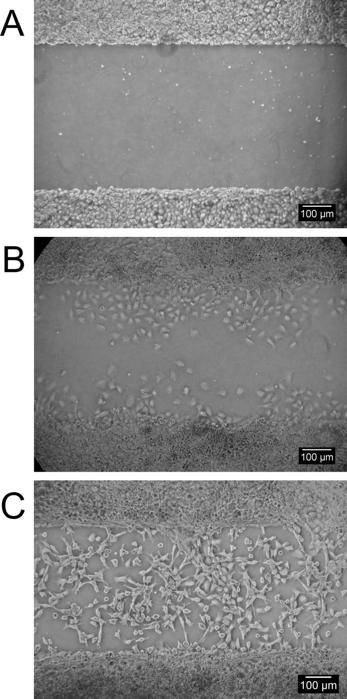
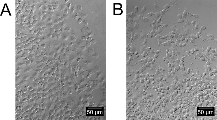
Quantifying the ability of a compound to modulate cell migration rate is a crucial part of many studies including those on chemotaxis, wound healing and cancer metastasis. Existing migration assays all have their strengths and weaknesses. The "scratch" assay is the most widely used because it seems appealingly simple and inexpensive. However, the scratch assay has some important limitations, as the tool introducing the "wound" might injure/stress the boundary cells and/or harm underlying matrix coatings, which in both cases will affect cell migration. This described method is a Cell Exclusion Zone Assay, in which cell-free areas are created by growing cells around removable silicone stoppers. Upon appropriate staining with fluorescent dyes and microscopically visualizing the monolayers, the migration rate is then quantified by counting the cells (nuclei) intruding the void area left by the silicone insert. In the current study human small intestine epithelial cells were seeded on a physiological substrate matrix to produce collectively migrating monolayers. Different substrates were tested to determine the optimal surface for enterocyte adherence and migration and morphological changes monitored. Recombinant human epidermal growth factor and osteopontin purified from urine were tested to see if the established migration assay produces accurate and reliable migration data with human small intestine cells. The obtained data accurately confirmed that the two bioactive proteins modulate cellular migration in a dose-dependent manner. The presented assay can likely be converted for use with other adherent cell lines or substrate matrices and allows for high throughput, while cost is kept low and versatility high. Co-staining can be applied in order to assay for cell death, different cell types, cell stress and others allowing intricate analysis of migration rate of mixed populations and correction for cell viability.
量化一种化合物调节细胞迁移速率的能力是许多研究的关键部分,包括那些关于趋化性、伤口愈合和癌症转移的研究。现有的迁移分析方法都有其优缺点。“划痕”分析是使用最广泛的,因为它看起来简单且成本低。然而,划痕分析有一些重要的局限性,因为引入“伤口”的工具可能会损伤/刺激边界细胞和/或损害底层的基质涂层,在这两种情况下都会影响细胞迁移。本文所述的方法是细胞排除区分析,其中通过在可移除的硅胶塞周围培养细胞来创建无细胞区域。在用荧光染料进行适当染色并在显微镜下观察单层细胞后,通过计数侵入硅胶插入物留下的空白区域的细胞(细胞核)来量化迁移速率。在当前研究中,将人小肠上皮细胞接种在生理底物基质上以产生集体迁移的单层细胞。测试了不同的底物以确定肠上皮细胞粘附和迁移的最佳表面,并监测形态变化。测试了从尿液中纯化的重组人表皮生长因子和骨桥蛋白,以确定所建立的迁移分析是否能产生关于人小肠细胞的准确可靠的迁移数据。获得的数据准确地证实了这两种生物活性蛋白以剂量依赖性方式调节细胞迁移。所提出的分析方法可能可以转换用于其他贴壁细胞系或底物基质,并允许高通量,同时成本保持较低且通用性较高。可以应用共染色来检测细胞死亡、不同细胞类型、细胞应激等,从而对混合群体的迁移速率进行复杂分析并校正细胞活力。