Zhou Yan, Shi Jianhua, Chu Dandan, Hu Wen, Guan Zongyu, Gong Cheng-Xin, Iqbal Khalid, Liu Fei
Key Laboratory of Neuroregeneration of Jiangsu and Ministry of Education of China, Co-innovation Center of Neuroregeneration, Nantong University, Nantong, China.
Department of Neurochemistry, Inge Grundke-Iqbal Research Floor, New York State Institute for Basic Research in Developmental Disabilities, New York, NY, United States.
Front Aging Neurosci. 2018 Feb 6;10:27. doi: 10.3389/fnagi.2018.00027. eCollection 2018.
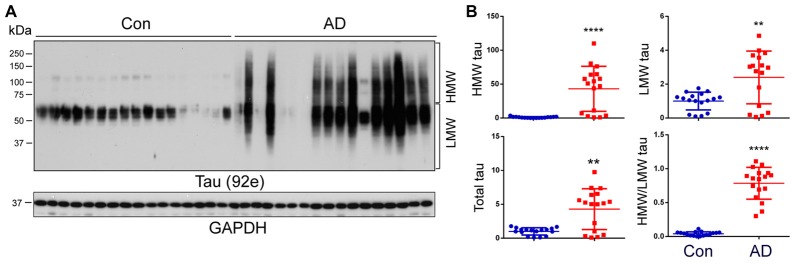
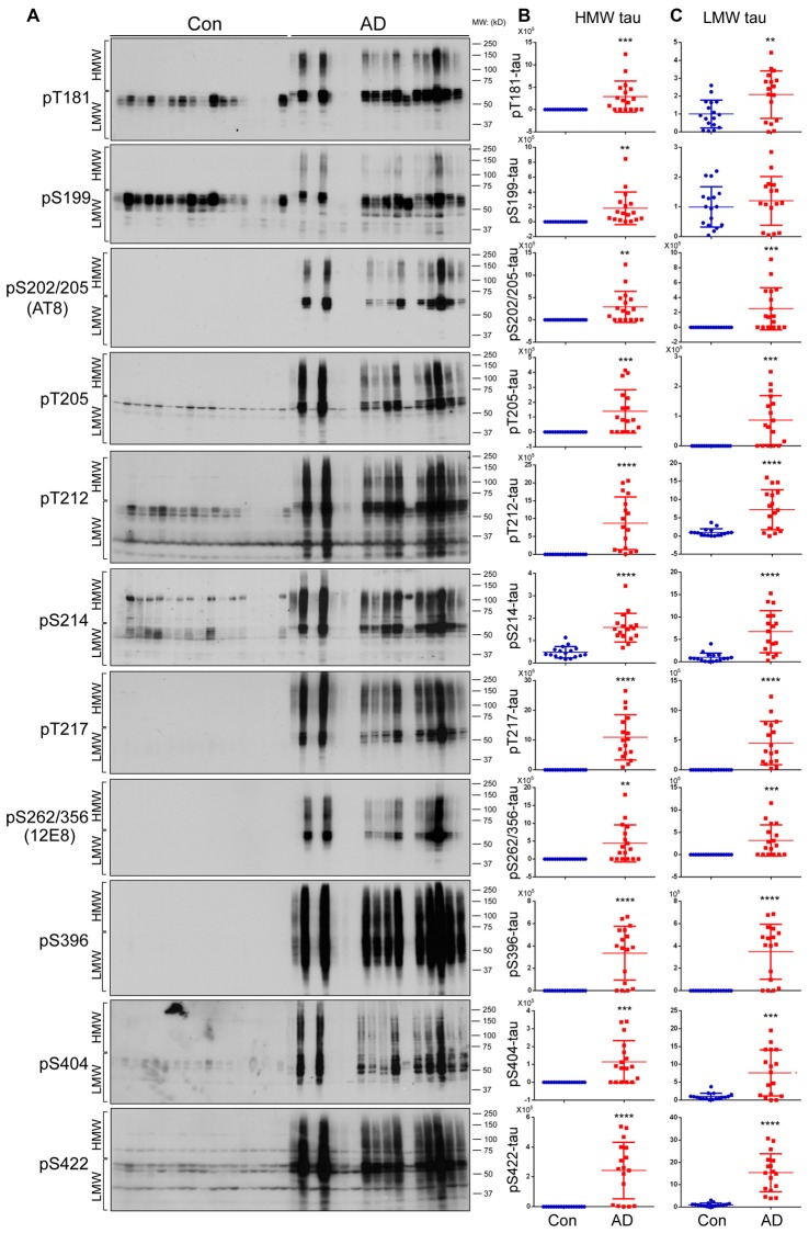
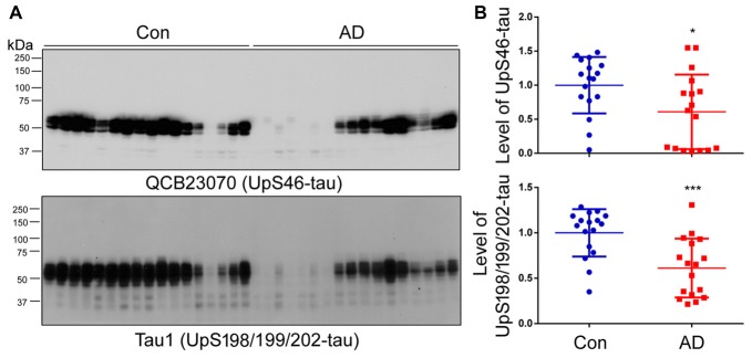
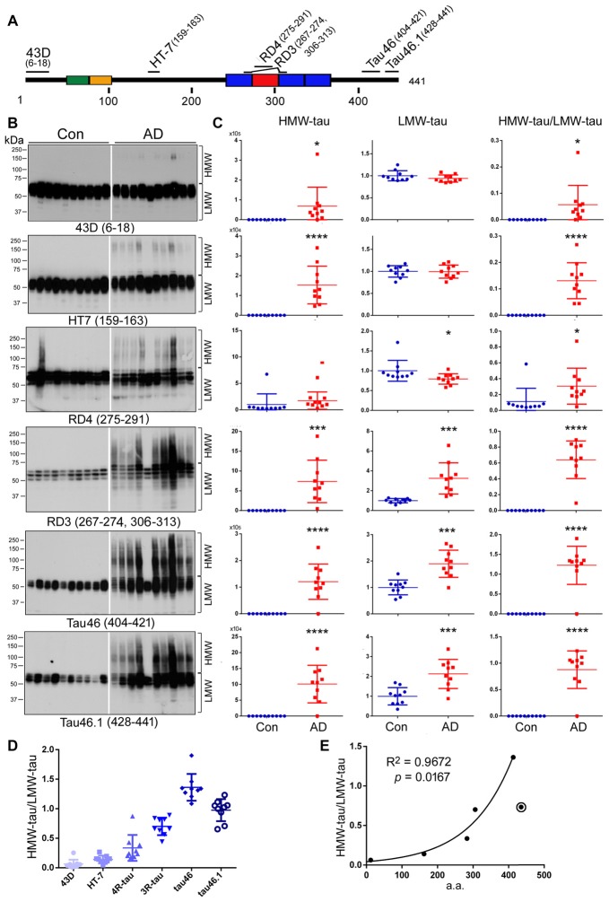
Microtubule (MT) associated protein tau is abnormally hyperphosphorylated and aggregated into paired helical filaments (PHFs), which manifest as neurofibrillary tangles (NFTs) in the brains of individuals with Alzheimer's disease (AD) and related tauopathies. Hyperphosphorylation and truncation of tau have been linked to the progression of the disease. However, the nature of phosphorylation and truncation of tau in AD brain are not very clear. In the present study we investigated the association of phosphorylation and truncation with high-molecular weight oligomers of tau (HMW-tau) in post-mortem AD brain by western blots. We found that tau from AD brain appears as a smear from low molecular weight (LMW) to HMW tau species in western blots developed with pan-tau antibodies. Similar level of LMW-tau was found in AD and control brains, whereas HMW-tau was found in AD brain only. HMW-tau was hyperphosphorylated at multiple sites and not unphosphorylated at Ser46 or Ser198/199/202. HMW-tau was weakly labeled by tau antibodies 43D against a.a. 6-18 and HT7 against a.a. 159-163 of tau, whereas, the C-terminal antibodies, tau46 and tau46.1, strongly labeled HMW-tau. The ratio of HMW-tau/LMW-tau detected by tau antibodies increased as the epitope of the tau antibodies ranges from N-terminal to C-terminal. The level of tau truncated at Asp421 was increased in AD brain, but was poorly associated with the HMW-tau. These findings suggest that tau pathogenesis involves both hyperphosphorylation and dominantly N-terminal truncation of tau in AD.
微管相关蛋白tau异常高度磷酸化并聚集成双螺旋丝(PHF),在阿尔茨海默病(AD)及相关tau蛋白病患者的大脑中表现为神经原纤维缠结(NFT)。tau蛋白的过度磷酸化和截短与疾病进展有关。然而,AD大脑中tau蛋白磷酸化和截短的本质尚不完全清楚。在本研究中,我们通过蛋白质印迹法研究了死后AD大脑中tau蛋白的磷酸化和截短与高分子量tau寡聚体(HMW-tau)的关系。我们发现,在用泛tau抗体进行蛋白质印迹时,AD大脑中的tau蛋白在低分子量(LMW)到HMW tau种类之间呈现为一条拖尾条带。在AD大脑和对照大脑中发现的LMW-tau水平相似,而HMW-tau仅在AD大脑中发现。HMW-tau在多个位点高度磷酸化,在Ser46或Ser198/199/202处未磷酸化。HMW-tau被针对tau蛋白第6 - 18位氨基酸的tau抗体43D和针对tau蛋白第159 - 163位氨基酸的HT7弱标记,而C末端抗体tau46和tau46.1则强烈标记HMW-tau。随着tau抗体的表位从N末端到C末端,tau抗体检测到的HMW-tau/LMW-tau比值增加。在AD大脑中,在Asp421处截短的tau蛋白水平升高,但与HMW-tau的相关性较差。这些发现表明,在AD中tau蛋白致病机制涉及tau蛋白的过度磷酸化和主要是N末端截短。