Department of Genetics and Microbiology, Universitat Autònoma de Barcelona, Barcelona, Spain.
Centre of Excellence in Experimental and Computational Developmental Biology, Institute of Biotechnology, University of Helsinki, Helsinki, Finland.
PLoS Comput Biol. 2018 Feb 26;14(2):e1005981. doi: 10.1371/journal.pcbi.1005981. eCollection 2018 Feb.
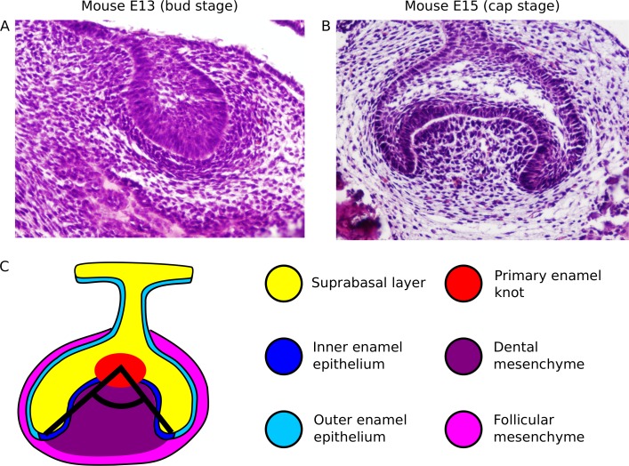
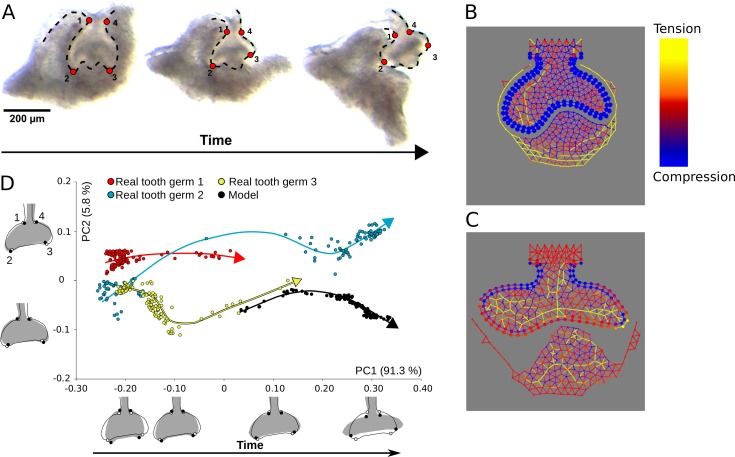
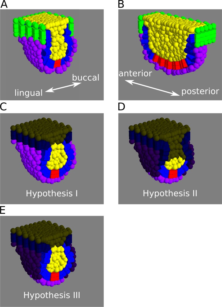
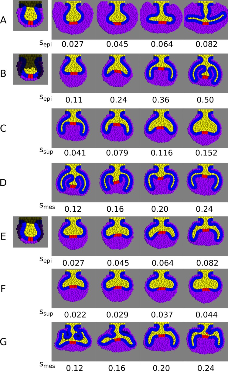
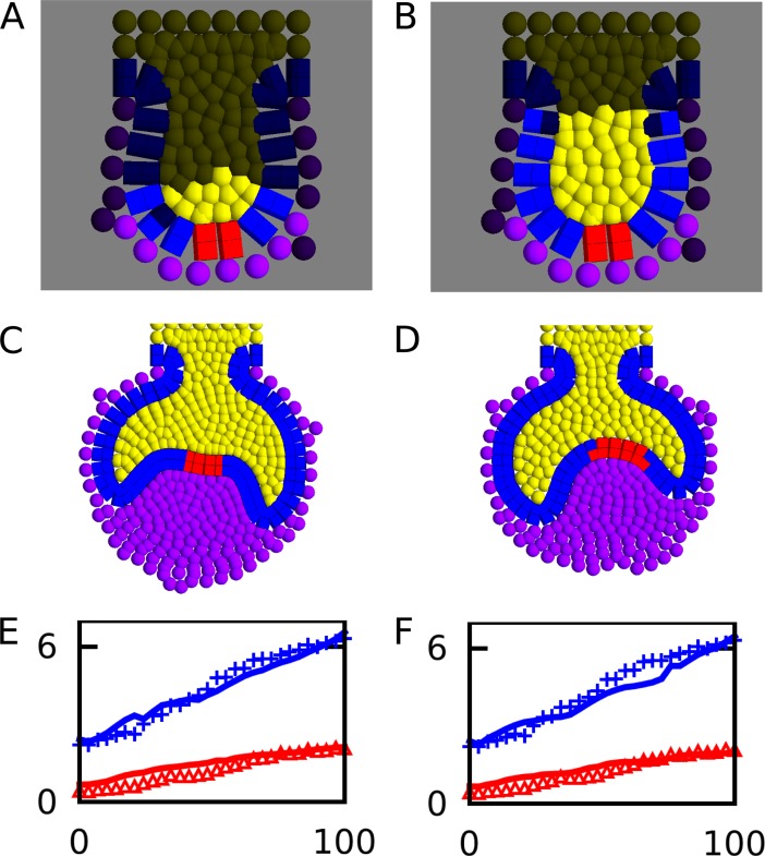
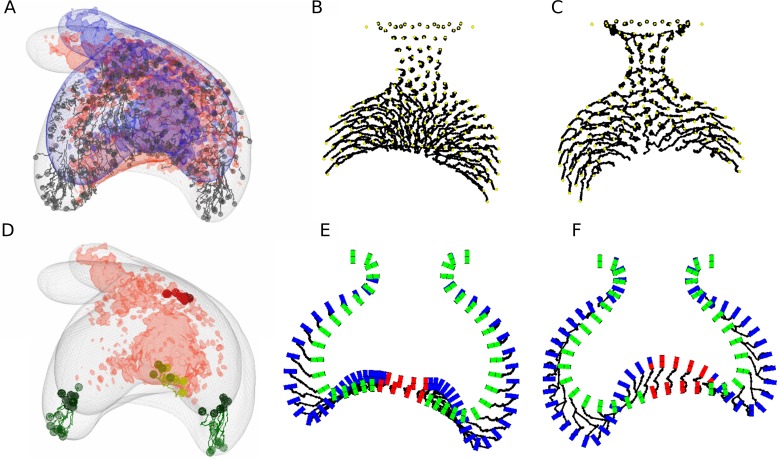
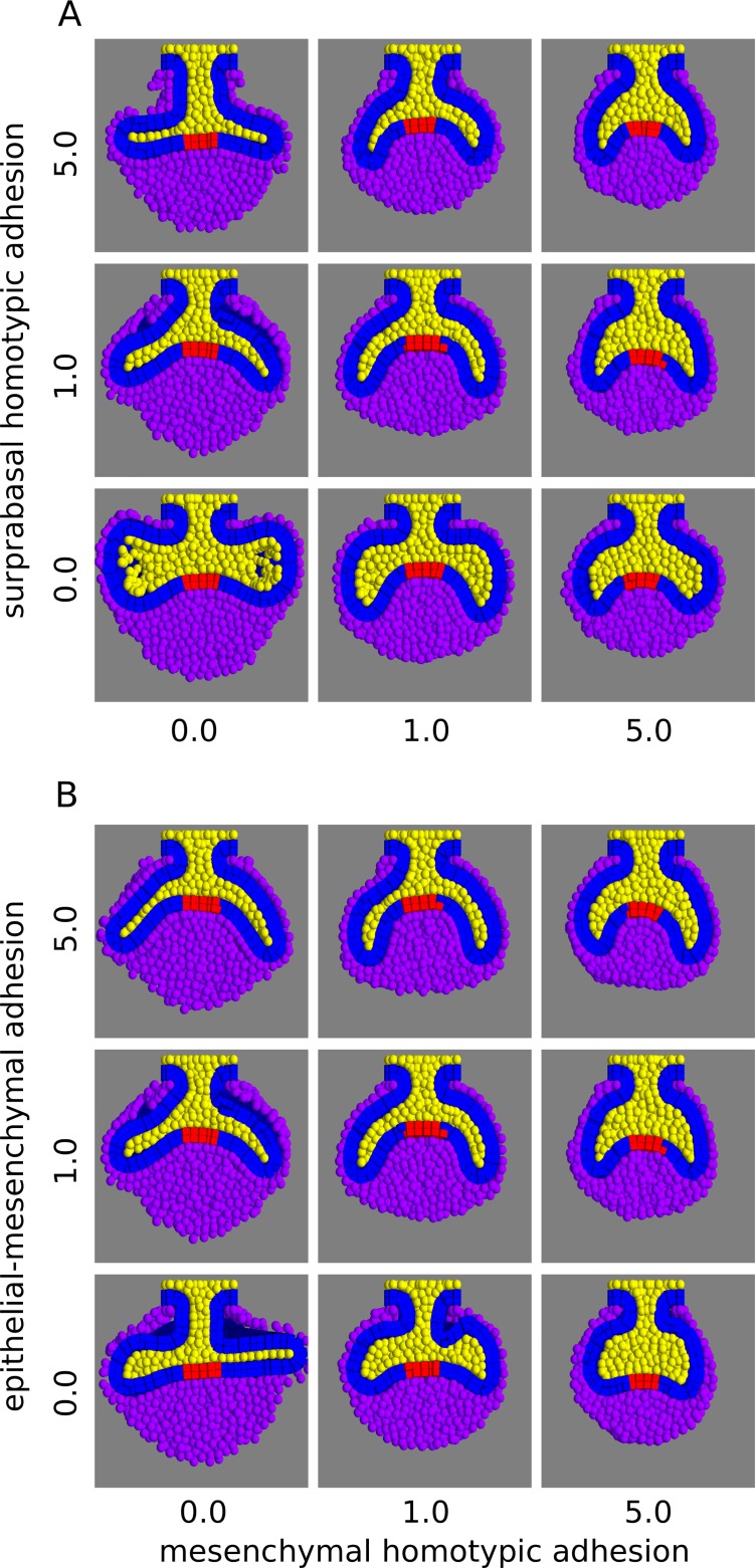
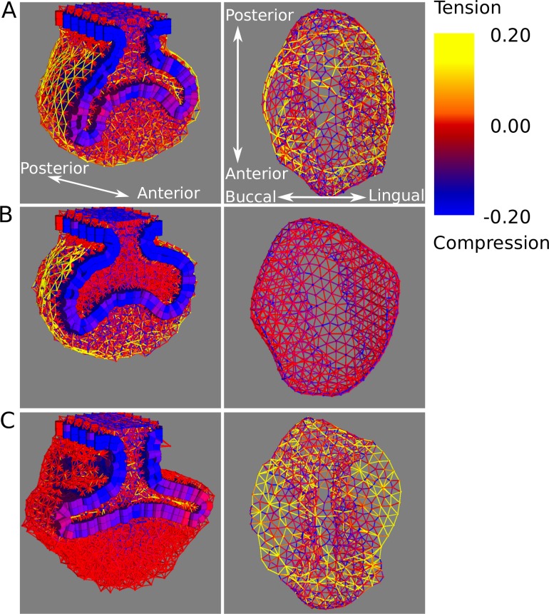
From gastrulation to late organogenesis animal development involves many genetic and bio-mechanical interactions between epithelial and mesenchymal tissues. Ectodermal organs, such as hairs, feathers and teeth are well studied examples of organs whose development is based on epithelial-mesenchymal interactions. These develop from a similar primordium through an epithelial folding and its interaction with the mesenchyme. Despite extensive knowledge on the molecular pathways involved, little is known about the role of bio-mechanical processes in the morphogenesis of these organs. We propose a simple computational model for the biomechanics of one such organ, the tooth, and contrast its predictions against cell-tracking experiments, mechanical relaxation experiments and the observed tooth shape changes over developmental time. We found that two biomechanical processes, differential tissue growth and differential cell adhesion, were enough, in the model, for the development of the 3D morphology of the early tooth germ. This was largely determined by the length and direction of growth of the cervical loops, lateral folds of the enamel epithelium. The formation of these cervical loops was found to require accelerated epithelial growth relative to other tissues and their direction of growth depended on specific differential adhesion between the three tooth tissues. These two processes and geometrical constraints in early tooth bud also explained the shape asymmetry between the lateral cervical loops and those forming in the anterior and posterior of the tooth. By performing mechanical perturbations ex vivo and in silico we inferred the distribution and direction of tensile stresses in the mesenchyme that restricted cervical loop lateral growth and forced them to grow downwards. Overall our study suggests detailed quantitative explanations for how bio-mechanical processes lead to specific morphological 3D changes over developmental time.
从原肠胚形成到晚期器官发生,动物发育涉及上皮组织和间充质组织之间的许多遗传和生物力学相互作用。外胚层器官,如毛发、羽毛和牙齿,是发育基于上皮-间充质相互作用的器官的良好研究范例。这些器官从类似的原基发育而来,通过上皮折叠及其与间充质的相互作用。尽管对涉及的分子途径有广泛的了解,但对于生物力学过程在这些器官形态发生中的作用知之甚少。我们提出了一个用于牙齿等器官生物力学的简单计算模型,并将其预测与细胞跟踪实验、力学松弛实验以及观察到的牙齿形状随时间的变化进行对比。我们发现,在模型中,两种生物力学过程,即组织差异生长和细胞差异粘附,足以发展早期牙胚的 3D 形态。这在很大程度上取决于颈环的生长长度和方向,颈环是釉质上皮的侧向褶皱。发现这些颈环的形成需要相对于其他组织加速上皮生长,并且它们的生长方向取决于三个牙齿组织之间的特定差异粘附。这两个过程和早期牙蕾中的几何约束也解释了侧向颈环与在牙齿前后形成的颈环之间的形状不对称性。通过进行离体和计算机模拟的机械扰动,我们推断出限制颈环侧向生长并迫使它们向下生长的间质中拉伸应力的分布和方向。总的来说,我们的研究表明,生物力学过程如何导致特定的形态 3D 变化在发育时间。