Department of Radiation Oncology, Technical University of Munich (TUM), Munich, Germany.
Department of Urology, Technical University Munich (TUM), Munich, Germany.
Radiat Oncol. 2018 Mar 1;13(1):36. doi: 10.1186/s13014-018-0977-2.
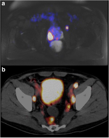
Ga-PSMA-PET-imaging has proven to be a highly sensitive and specific diagnostic element for patients with prostate cancer (PC). Does the standard clinical target volume (CTV) cover the majority of Ga-PSMA-PET detected lymph nodes (LNs) in a primary setting?
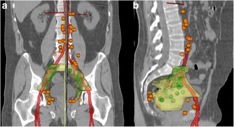
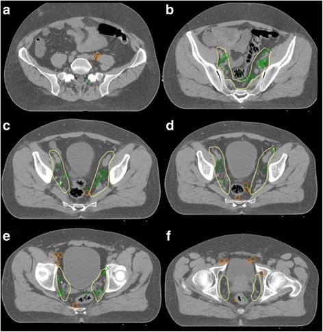
25 out of 159 patients with primary PC who underwent Ga-PSMA-PET-imaging were analyzed in the process of this study. These 25 high-risk patients had a total of 126 LNs with positive Ga-PSMA-ligand uptake. A standard CTV according to the 'Radiation Therapy Oncology Group' consensus was delineated and LNs were judged whether they were in- or outside of this target volume. With a Pearson correlation we additionally evaluated whether the Gleason score, the prostate-specific antigen (PSA) value or the risk according to the Roach formula correlate with a higher chance of LNs being outside of the CTV in uncommon LN locations.
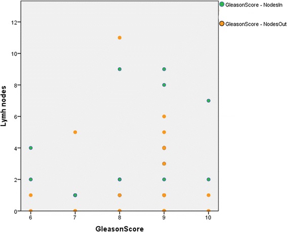
81 (64.3%) of 126 LNs were covered by the CTV with a complete coverage of all positive LNs inside the respective radiation volume in 11 of 25 patients (44%). LNs that were not covered by the CTV included (para-aortic,) common-iliac, pre-sacral, obturatoric, para-rectal, para-vesical and pre-acetabular locations. In a statistical analysis neither the Gleason score, nor the PSA value, nor the calculated risk with the Roach formula correlated with LNs being inside or outside of the CTV in this patient group.
Ga-PSMA-PET-imaging proves to be a valuable asset for patients and physicians for primary diagnosis and treatment planning. In our study, trusting the RTOG consensus for CTV delineation would have led to up to 35.7% of all LNs not to be included in the clinical radiation volume, which might have resulted in insufficient radiation dose coverage.
镓-PSMA-PET 成像已被证明是前列腺癌(PC)患者高度敏感和特异的诊断手段。在原发情况下,标准临床靶区(CTV)是否能涵盖大多数镓-PSMA-PET 检测到的淋巴结(LN)?
在这项研究中,分析了 159 例接受镓-PSMA-PET 成像的原发性 PC 患者中的 25 例。这 25 例高危患者共有 126 个 LN 摄取阳性的 Ga-PSMA 配体。根据“放射治疗肿瘤学组”共识,划定了标准 CTV,并判断 LN 是否位于该靶区内外。通过皮尔逊相关性分析,我们还评估了 Gleason 评分、前列腺特异性抗原(PSA)值或 Roach 公式风险是否与 LN 位于不常见 LN 位置的 CTV 外的可能性更高相关。
126 个 LN 中有 81 个(64.3%)被 CTV 覆盖,在 25 例患者中的 11 例(44%)中,所有阳性 LN 均完全位于相应放射区域内。未被 CTV 覆盖的 LN 包括(腹主动脉旁、髂总、骶前、闭孔、直肠旁、膀胱旁和髋臼前)。在统计学分析中,Gleason 评分、PSA 值或 Roach 公式计算的风险均与该患者组中 LN 位于 CTV 内或外无关。
镓-PSMA-PET 成像对患者和医生进行原发性诊断和治疗计划非常有价值。在我们的研究中,仅依赖 RTOG 共识进行 CTV 勾画可能导致多达 35.7%的 LN 不包括在临床照射体积中,这可能导致照射剂量覆盖不足。