Anwar Adeel, Zhang Zhen, Lv Decheng, Lv Gang, Zhao Zhi, Wang Yanfeng, Cai Yue, Qasim Wasim, Nazir Muhammad Umar, Lu Ming
Department of Orthopaedic Surgery, The First Affiliated Hospital of Dalian Medical University, 222 Zhongshan road, 116011, Dalian, Liaoning, People's Republic of China.
Department of Orthopaedic Surgery, The First Affiliated Hospital of China Medical University, 155 Nanjing north street, 110001, Shenyang, Liaoning, People's Republic of China.
BMC Musculoskelet Disord. 2018 Mar 6;19(1):73. doi: 10.1186/s12891-018-1989-7.
Clinically there are different fixation methods used for fixation of the posterior malleolar fractures (PMF), but the best treatment modality is still not clear. Few studies have concentrated on this issue, least of all using a biomechanical comparison. The purpose of this study was to carry out a computational comparative biomechanics of three different commonly used fixation constructs for the fixation of PMF by finite element analysis (FEA).
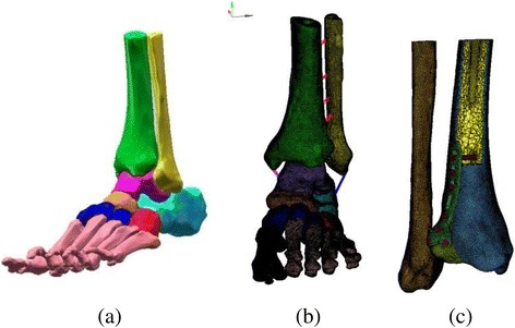
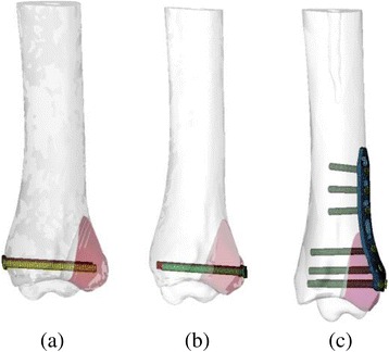
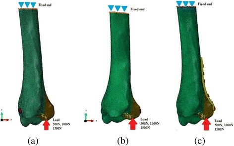
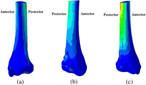
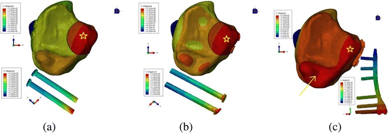
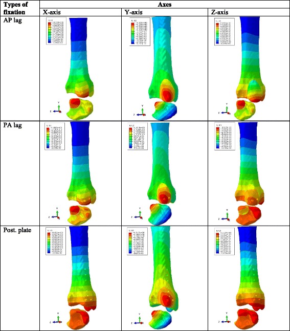
Computed tomography (CT) images were used to reconstruct three dimensional (3D) model of the tibia. Computer aided design (CAD) software was used to design 3D models of PMF. Finally, 3D models of PMF fixed with two antero-posterior (AP) lag screws, two postero-anterior (PA) lag screws and posterior plate were simulated through computational processing. Simulated loads of 500 N, 1000 N and 1500 N were applied to the PMF and proximal ends of the models were fixed in all degrees of freedom. Output results representing the model von Mises stress, relative fracture micro-motion and vertical displacement of the fracture fragment were analyzed.
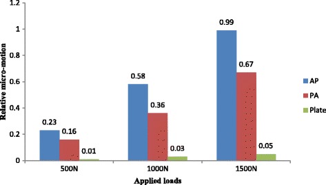
The mean vertical displacement value in the posterior plate group (0.52 mm) was lower than AP (0.68 mm) and PA (0.69 mm) lag groups. Statistically significant low amount of the relative micro-motion (P < 0.05) was observed in the posterior plate group.
It was concluded that the posterior plate is biomechanically the most stable fixation method for fixation of PMF.
临床上用于固定后踝骨折(PMF)的方法各异,但最佳治疗方式仍不明确。很少有研究关注这个问题,尤其是进行生物力学比较的研究。本研究的目的是通过有限元分析(FEA)对三种不同的常用固定结构用于PMF固定进行计算比较生物力学研究。
使用计算机断层扫描(CT)图像重建胫骨的三维(3D)模型。利用计算机辅助设计(CAD)软件设计PMF的3D模型。最后,通过计算处理模拟用两枚前后(AP)拉力螺钉、两枚后前(PA)拉力螺钉和后外侧钢板固定的PMF的3D模型。对PMF施加500 N、1000 N和1500 N的模拟载荷,并将模型近端的所有自由度固定。分析代表模型冯·米塞斯应力、相对骨折微动和骨折块垂直位移的输出结果。
后外侧钢板组的平均垂直位移值(0.52 mm)低于AP(0.68 mm)和PA(0.69 mm)拉力螺钉组。后外侧钢板组观察到相对微动的量在统计学上显著较低(P < 0.05)。
得出结论,后外侧钢板在生物力学上是固定PMF最稳定的固定方法。