Mechelinck Mare, Hein Marc, Bellen Sven, Rossaint Rolf, Roehl Anna B
Department of Anesthesiology, Medical Faculty, RWTH Aachen University, Aachen, Germany.
Physiol Rep. 2018 Mar;6(5). doi: 10.14814/phy2.13605.
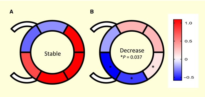
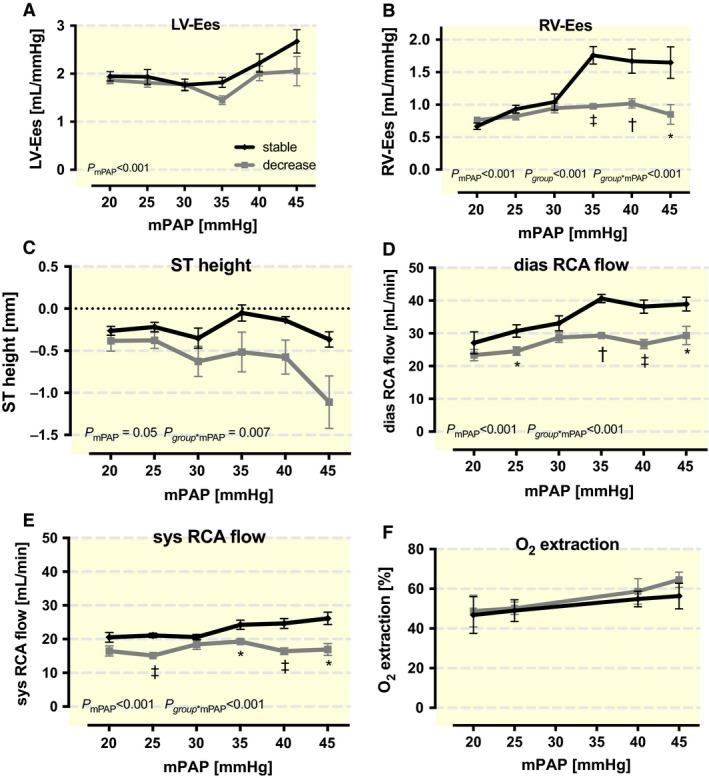
The extent of right ventricular compensation compared to the left ventricle is restricted and varies among individuals, which makes it difficult to define. While establishing a model of acute pulmonary hypertension in pigs we observed two different kinds of compensation in our animals. Looking deeper into the hemodynamic data we tried to delineate why some animals could compensate and others could not. Pulmonary hypertension (mean pressure 45 mmHg) was induced gradually by infusion of a stable thromboxane A analogue U46619 in a porcine model (n = 22). Hemodynamic data (pressure-volume loops, strain-analysis of echocardiographic data and coronary flow measurements) were evaluated retrospectively for the short-term right ventricular compensatory mechanisms and limits (Roehl et al. [2012] Acta Anaesthesiol. Scand., 56:449-58) 10 animals showed stable arterial blood pressures, whereas 12 pigs exhibited a significant drop of 16.4 ± 9.9 mmHg. Cardiac output and heart rate were comparable in both groups. In contrast, right ventricular contractility and coronary flow only rose in the stable group. The unchanging values in the decrease group correlated with an increasing ST-segment depression and a loss of ventricular synchronism and resulted in a larger septum bulging to the right ventricle. Simultaneously, a reduced left-ventricular end-diastolic volume and a missing improvement in contractility in the posterior septal and inferior free wall of the left ventricle have been observed. Our findings suggest that right ventricular compensation during acute pulmonary hypertension is strongly dependent on the individual capability to increase coronary flow. The cause for inter-individual variability could be the dimension and reactivity of the coronary system.
与左心室相比,右心室的代偿程度有限且个体差异较大,这使得其难以界定。在建立猪急性肺动脉高压模型的过程中,我们观察到实验动物出现了两种不同类型的代偿情况。深入研究血流动力学数据后,我们试图弄清楚为何有些动物能够代偿而有些则不能。通过在猪模型(n = 22)中逐渐输注稳定的血栓素A类似物U46619来诱导肺动脉高压(平均压45 mmHg)。对血流动力学数据(压力-容积环、超声心动图数据的应变分析以及冠状动脉血流测量)进行回顾性评估,以研究短期右心室的代偿机制及限度(Roehl等人,[2012]《麻醉学与复苏杂志》,56:449 - 58)。10只动物的动脉血压保持稳定,而12只猪的动脉血压显著下降了16.4 ± 9.9 mmHg。两组的心输出量和心率相当。相比之下,只有稳定组的右心室收缩力和冠状动脉血流有所增加。血压下降组中各项指标未发生变化,这与ST段压低增加、心室同步性丧失相关,并导致室间隔向右心室凸出增大。同时,还观察到左心室舒张末期容积减小,且左心室后间隔和下壁的收缩力未得到改善。我们的研究结果表明,急性肺动脉高压期间右心室的代偿很大程度上取决于个体增加冠状动脉血流的能力。个体差异的原因可能是冠状动脉系统的大小和反应性。