• 文献检索
  • 文档翻译
  • 深度研究
  • 学术资讯
  • Suppr Zotero 插件Zotero 插件
  • 邀请有礼
  • 套餐&价格
  • 历史记录
应用&插件
Suppr Zotero 插件Zotero 插件浏览器插件Mac 客户端Windows 客户端微信小程序
定价
高级版会员购买积分包购买API积分包
服务
文献检索文档翻译深度研究API 文档MCP 服务
关于我们
关于 Suppr公司介绍联系我们用户协议隐私条款
关注我们

Suppr 超能文献

核心技术专利:CN118964589B侵权必究
粤ICP备2023148730 号-1Suppr @ 2026

文献检索

告别复杂PubMed语法,用中文像聊天一样搜索,搜遍4000万医学文献。AI智能推荐,让科研检索更轻松。

立即免费搜索

文件翻译

保留排版,准确专业,支持PDF/Word/PPT等文件格式,支持 12+语言互译。

免费翻译文档

深度研究

AI帮你快速写综述,25分钟生成高质量综述,智能提取关键信息,辅助科研写作。

立即免费体验

继发性高血压患者的多模态影像学检查:重点关注根据病因选择合适的影像学检查方法。

Multimodality Imaging in Patients with Secondary Hypertension: With a Focus on Appropriate Imaging Approaches Depending on the Etiologies.

机构信息

Department of Radiology, Seoul National University Bundang Hospital, Seongnam 13620, Korea.

Department of Internal Medicine, Seoul National University Bundang Hospital, Seongnam 13620, Korea.

出版信息

Korean J Radiol. 2018 Mar-Apr;19(2):272-283. doi: 10.3348/kjr.2018.19.2.272. Epub 2018 Feb 22.

DOI:10.3348/kjr.2018.19.2.272
PMID:29520185
原文链接:https://pmc.ncbi.nlm.nih.gov/articles/PMC5840056/
Abstract

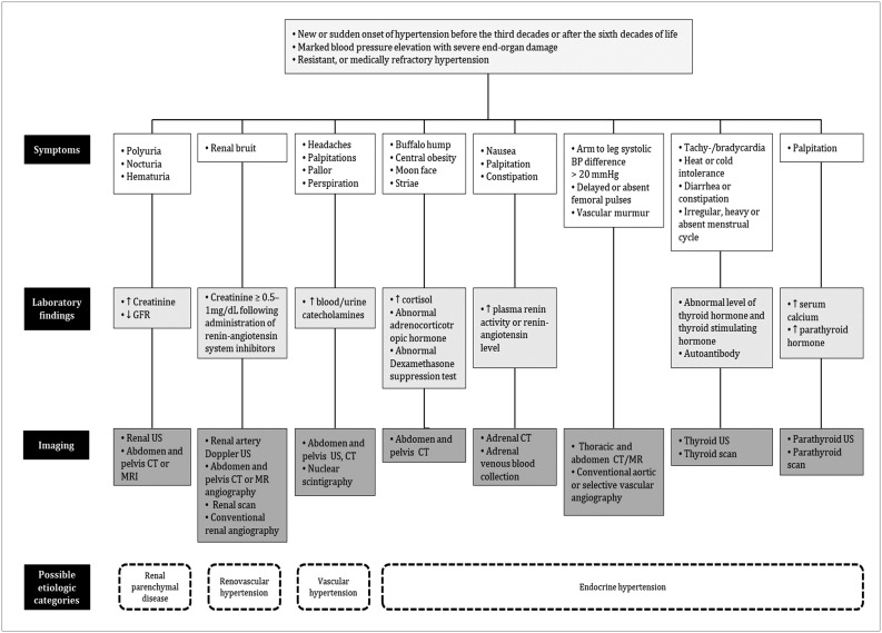
Although the causes of hypertension are usually unknown, about 10% of the cases occur secondary to specific etiologies, which are often treatable. Common categories of secondary hypertension include renal parenchymal disease, renovascular stenosis, vascular and endocrinologic disorders. For diseases involving the renal parenchyma and adrenal glands, ultrasonography (US), computed tomography (CT) or magnetic resonance (MR) imaging is recommended. For renovascular stenosis and vascular disorders, Doppler US, conventional or noninvasive (CT or MR) angiography is an appropriate modality. Nuclear imaging can be useful in the differential diagnosis of endocrine causes. Radiologists should understand the role of each imaging modality and its typical findings in various causes of secondary hypertension. This article focuses on appropriate imaging approaches in accordance with the categorized etiologies leading to hypertension.

摘要

虽然高血压的病因通常不明确,但约 10%的病例继发于特定病因,这些病因通常是可治疗的。继发性高血压的常见类型包括肾实质疾病、肾血管性狭窄、血管和内分泌疾病。对于涉及肾脏实质和肾上腺的疾病,推荐使用超声检查(US)、计算机断层扫描(CT)或磁共振成像(MR)。对于肾血管性狭窄和血管疾病,多普勒 US、传统或无创(CT 或 MR)血管造影是合适的方式。核医学成像在鉴别内分泌原因方面具有一定作用。放射科医生应了解每种成像方式的作用及其在继发性高血压各种病因中的典型表现。本文重点介绍了根据导致高血压的分类病因选择合适的影像学方法。

https://cdn.ncbi.nlm.nih.gov/pmc/blobs/a2b5/5840056/aafa8d852000/kjr-19-272-g012.jpg
https://cdn.ncbi.nlm.nih.gov/pmc/blobs/a2b5/5840056/07dce42850ac/kjr-19-272-g001.jpg
https://cdn.ncbi.nlm.nih.gov/pmc/blobs/a2b5/5840056/c88cdf3eb1f2/kjr-19-272-g002.jpg
https://cdn.ncbi.nlm.nih.gov/pmc/blobs/a2b5/5840056/8ddb66559ebc/kjr-19-272-g003.jpg
https://cdn.ncbi.nlm.nih.gov/pmc/blobs/a2b5/5840056/1ff1a98a3c7e/kjr-19-272-g004.jpg
https://cdn.ncbi.nlm.nih.gov/pmc/blobs/a2b5/5840056/269bab5729a7/kjr-19-272-g005.jpg
https://cdn.ncbi.nlm.nih.gov/pmc/blobs/a2b5/5840056/fd175413721b/kjr-19-272-g006.jpg
https://cdn.ncbi.nlm.nih.gov/pmc/blobs/a2b5/5840056/d9e32da9d00f/kjr-19-272-g007.jpg
https://cdn.ncbi.nlm.nih.gov/pmc/blobs/a2b5/5840056/3648f481b26a/kjr-19-272-g008.jpg
https://cdn.ncbi.nlm.nih.gov/pmc/blobs/a2b5/5840056/b768e7c650f5/kjr-19-272-g009.jpg
https://cdn.ncbi.nlm.nih.gov/pmc/blobs/a2b5/5840056/6724696d40ec/kjr-19-272-g010.jpg
https://cdn.ncbi.nlm.nih.gov/pmc/blobs/a2b5/5840056/fea1f602058d/kjr-19-272-g011.jpg
https://cdn.ncbi.nlm.nih.gov/pmc/blobs/a2b5/5840056/aafa8d852000/kjr-19-272-g012.jpg

相似文献

1
Multimodality Imaging in Patients with Secondary Hypertension: With a Focus on Appropriate Imaging Approaches Depending on the Etiologies.继发性高血压患者的多模态影像学检查:重点关注根据病因选择合适的影像学检查方法。
Korean J Radiol. 2018 Mar-Apr;19(2):272-283. doi: 10.3348/kjr.2018.19.2.272. Epub 2018 Feb 22.
2
Imaging modalities for renal artery stenosis in suspected renovascular hypertension: prospective intraindividual comparison of color Doppler US, CT angiography, GD-enhanced MR angiography, and digital substraction angiography.疑似肾血管性高血压患者肾动脉狭窄的成像方式:彩色多普勒超声、CT血管造影、钆增强磁共振血管造影和数字减影血管造影的前瞻性个体内比较
Ren Fail. 2007;29(3):295-302. doi: 10.1080/08860220601166305.
3
[Rational diagnostic approach in hypertension].[高血压的合理诊断方法]
Praxis (Bern 1994). 2003 Dec 3;92(49):2087-93. doi: 10.1024/0369-8394.92.49.2087.
4
New Insights Into Pathophysiology, Diagnosis, and Treatment of Renovascular Hypertension.肾血管性高血压病理生理学、诊断及治疗的新见解
Iran J Kidney Dis. 2017 Mar;11(2):79-89.
5
Pediatric hypertension: an approach to imaging.小儿高血压:影像学检查方法
Pediatr Radiol. 1986;16(6):461-7. doi: 10.1007/BF02387958.
6
Renovascular hypertension. Radiographic methods.肾血管性高血压。影像学检查方法。
Q J Nucl Med. 2002 Dec;46(4):283-94.
7
Radiologic evaluation of suspected renovascular hypertension.疑似肾血管性高血压的放射学评估。
Am Fam Physician. 2009 Aug 1;80(3):273-9.
8
Renovascular hypertension.肾血管性高血压
Indian Pediatr. 2005 Jan;42(1):47-54.
9
Non-invasive imaging cannot replace formal angiography in the diagnosis of renovascular hypertension.在肾血管性高血压的诊断中,非侵入性成像不能替代正规的血管造影。
Pediatr Nephrol. 2017 Mar;32(3):495-502. doi: 10.1007/s00467-016-3501-7. Epub 2016 Oct 17.
10
Imaging of renovascular hypertension: respective values of renal scintigraphy, renal Doppler US, and MR angiography.肾血管性高血压的影像学检查:肾闪烁显像、肾多普勒超声及磁共振血管造影的各自价值
Radiographics. 2000 Sep-Oct;20(5):1355-68; discussion 1368-72. doi: 10.1148/radiographics.20.5.g00se131355.

引用本文的文献

1
A look at radiation detectors and their applications in medical imaging.探讨辐射探测器及其在医学成像中的应用。
Jpn J Radiol. 2024 Feb;42(2):145-157. doi: 10.1007/s11604-023-01486-z. Epub 2023 Sep 21.
2
Guideline for Cardiovascular Magnetic Resonance Imaging from the Korean Society of Cardiovascular Imaging-Part 1: Standardized Protocol.韩国心血管影像学会心血管磁共振成像指南-第 1 部分:标准化方案。
Korean J Radiol. 2019 Sep;20(9):1313-1333. doi: 10.3348/kjr.2019.0398.

本文引用的文献

1
Resistant hypertension caused by stenosis of the aorta in elderly women: three case reports.老年女性主动脉狭窄所致顽固性高血压:三例报告
Clin Hypertens. 2014 Nov 18;20:5. doi: 10.1186/s40885-014-0005-2. eCollection 2014.
2
Hypertension in Chronic Glomerulonephritis.慢性肾小球肾炎中的高血压
Electrolyte Blood Press. 2015 Dec;13(2):41-5. doi: 10.5049/EBP.2015.13.2.41. Epub 2015 Dec 30.
3
Coarctation of the aorta in adolescents and adults: A review of clinical features and CT imaging.青少年及成人主动脉缩窄:临床特征与CT影像综述
J Cardiovasc Comput Tomogr. 2016 Jan-Feb;10(1):1-12. doi: 10.1016/j.jcct.2015.11.002. Epub 2015 Nov 19.
4
Risk of Systemic Hypertension and Cerebrovascular Accident in Patients With Aortic Coarctation Aged <60 Years (from a National Database Study).60岁以下主动脉缩窄患者发生系统性高血压和脑血管意外的风险(来自一项全国性数据库研究)
Am J Cardiol. 2015 Sep 1;116(5):779-84. doi: 10.1016/j.amjcard.2015.05.052. Epub 2015 Jun 4.
5
CT and MR imaging for evaluation of cystic renal lesions and diseases.用于评估肾囊性病变和疾病的CT及磁共振成像。
Radiographics. 2015 Jan-Feb;35(1):125-41. doi: 10.1148/rg.351130016.
6
Characterization of lipid-poor adrenal adenoma: chemical-shift MRI and washout CT.乏脂性肾上腺腺瘤的特征:磁共振化学位移成像和 CT 廓清。
AJR Am J Roentgenol. 2014 May;202(5):1043-50. doi: 10.2214/AJR.13.11389.
7
Physiological basis for the etiology, diagnosis, and treatment of adrenal disorders: Cushing's syndrome, adrenal insufficiency, and congenital adrenal hyperplasia.肾上腺疾病的病因、诊断和治疗的生理学基础:库欣综合征、肾上腺功能不全和先天性肾上腺增生。
Compr Physiol. 2014 Apr;4(2):739-69. doi: 10.1002/cphy.c130035.
8
The Japanese Society of Hypertension Guidelines for the Management of Hypertension (JSH 2014).日本高血压学会高血压管理指南(JSH 2014)
Hypertens Res. 2014 Apr;37(4):253-390. doi: 10.1038/hr.2014.20.
9
Contrast-induced Nephropathy.对比剂肾病
Heart Views. 2013 Jul;14(3):106-16. doi: 10.4103/1995-705X.125926.
10
The thyroid: review of imaging features and biopsy techniques with radiologic-pathologic correlation.甲状腺:影像特征与活检技术综述及放射学与病理学相关性分析
Radiographics. 2014 Mar-Apr;34(2):276-93. doi: 10.1148/rg.342135067.