Assi Mohamad, Dauguet Nicolas, Jacquemin Patrick
de Duve Institute, Université catholique de Louvain, Brussels, Belgium.
de Duve Institute, Flow Cytometry and Cell Sorting Facility (CYTF), Brussels, Belgium.
Front Physiol. 2018 Feb 27;9:129. doi: 10.3389/fphys.2018.00129. eCollection 2018.
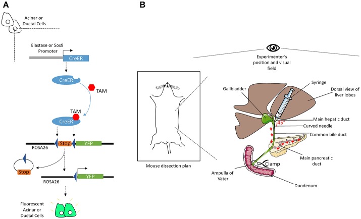
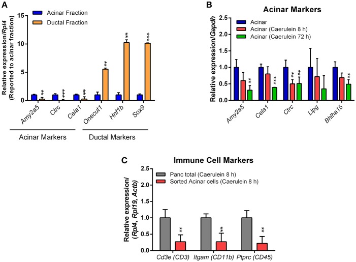
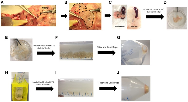
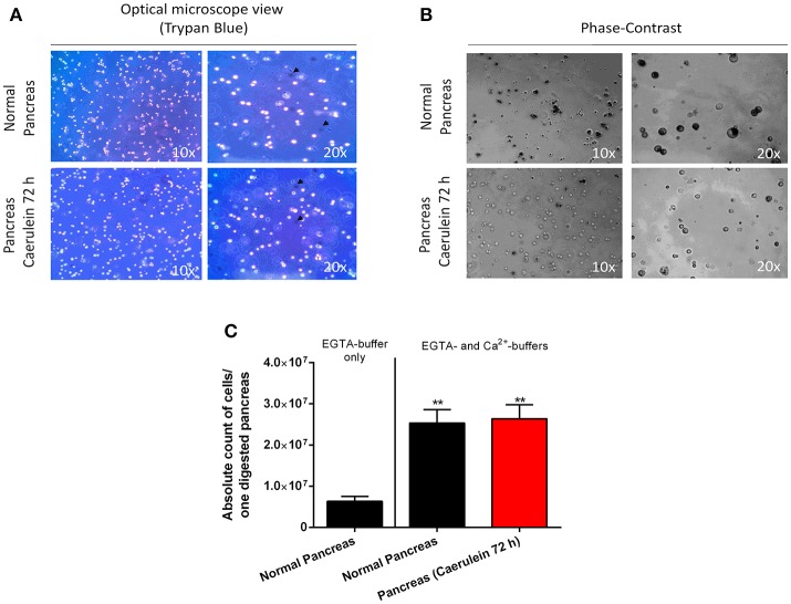
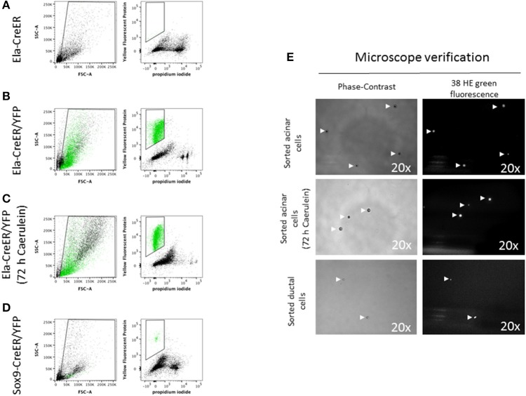
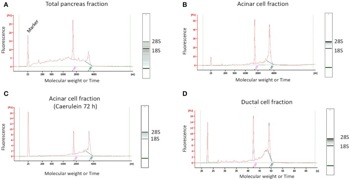
The isolation of ribonucleic acid (RNA) suitable for gene expression studies is challenging in the pancreas, due to its high ribonuclease activity. This is even more complicated during pancreatitis, a condition associated with inflammation and fibrosis. Our aim was to implement a time-effective and reproducible protocol to isolate high quality RNA from specific pancreatic cell subtypes, in normal and inflammatory conditions. We used two genetically engineered mouse models (GEMM), Ela-CreER/YFP and Sox9-CreER/YFP, to isolate acinar and ductal cells, respectively. To induce pancreatitis, mice received a caerulein treatment (125 μg/kg) for 8 and 72 h. We alternatively used EGTA and calcium buffers that contain collagenase P (0.6 mg/mL) to rapidly digest the pancreas into individual cells. Most of the cells from normal and injured pancreas were single-dissociated, exhibited a round morphology and did not incorporate trypan blue dye. Cell suspensions from Ela- and Sox9-CreER/YFP pancreas were then sorted by flow cytometry to isolate the YFP-positive acinar and ductal cells, respectively. Sorted cells kept a round shape and emitted fluorescence detected by the 38 HE green fluorescence filter. RNA was isolated by column-based purification approach. The RNA integrity number (RIN) was high in sorted acinar cell fractions treated with or without caerulein (8.6 ± 0.17 and 8.4 ± 0.09, respectively), compared to the whole pancreas fraction (4.8 ± 1.1). Given the low number of sorted ductal cells, the RIN value was slightly lower compared to acini (7.4 ± 0.4). Quantitative-PCR experiments indicated that sorted acinar and ductal cells express the specific acinar and ductal markers, respectively. Additionally, RNA preparations from caerulein-treated acinar cells were free from significant contamination with immune cell RNA. We thus validated the DIE (Digestion, Isolation, and Extraction)-RNA tool as a reproducible and efficient protocol to isolate pure acinar and ductal cells and to extract high quality RNA from these cells.
由于胰腺中核糖核酸酶(RNA)活性很高,因此分离适合基因表达研究的核糖核酸(RNA)具有挑战性。在胰腺炎(一种与炎症和纤维化相关的病症)期间,这一过程会更加复杂。我们的目标是实施一种省时且可重复的方案,以便在正常和炎症条件下从特定的胰腺细胞亚型中分离出高质量的RNA。我们使用了两种基因工程小鼠模型(GEMM),即Ela-CreER/YFP和Sox9-CreER/YFP,分别用于分离腺泡细胞和导管细胞。为了诱导胰腺炎,给小鼠注射雨蛙素(125μg/kg),持续8小时和72小时。我们还使用了含有胶原酶P(0.6mg/mL)的EGTA和钙缓冲液,将胰腺快速消化成单个细胞。来自正常和受损胰腺的大多数细胞都能被单个解离,呈现圆形形态,且不摄取台盼蓝染料。然后,通过流式细胞术对来自Ela-CreER/YFP和Sox9-CreER/YFP胰腺的细胞悬液进行分选,分别分离出YFP阳性的腺泡细胞和导管细胞。分选后的细胞保持圆形,并发出可被38 HE绿色荧光滤光片检测到的荧光。通过基于柱的纯化方法分离RNA。与整个胰腺部分(4.8±1.1)相比,用或不用雨蛙素处理的分选腺泡细胞部分的RNA完整性数值(RIN)较高(分别为8.6±0.17和8.4±0.09)。鉴于分选的导管细胞数量较少,其RIN值比腺泡细胞略低(7.4±0.4)。定量PCR实验表明,分选的腺泡细胞和导管细胞分别表达特定的腺泡和导管标志物。此外,来自雨蛙素处理的腺泡细胞的RNA制剂没有受到免疫细胞RNA的明显污染。因此,我们验证了DIE(消化、分离和提取)-RNA工具是一种可重复且高效的方案,用于分离纯净的腺泡细胞和导管细胞,并从这些细胞中提取高质量的RNA。