Bhogal Pervinder, AlMatter Muhammad, Hellstern Victoria, Ganslandt Oliver, Bäzner Hansjörg, Henkes Hans, Aguilar-Pérez Marta
Neuroradiological Clinic, Neurocenter, Klinikum Stuttgart, Germany.
Neurosurgical Clinic, Neurocenter, Klinikum Stuttgart, Germany.
Neurointervention. 2018 Mar;13(1):20-31. doi: 10.5469/neuroint.2018.13.1.20. Epub 2018 Mar 2.
The Medina Embolic Device (MED) is a new intrasaccular device with promising early results. Previously we documented our initial experience of this device both alone and in combination with other devices including flow diverter stents (FDS). We sought to determine the effect of the MED + FDS strategy for the treatment of selected aneurysms.
We performed a retrospective analysis of prospectively collected data to identify all patients with aneurysms treated using both the MED and intraluminal FDS. We present our technical success rate, early and mid-term angiographic follow-up, and clinical outcome data.
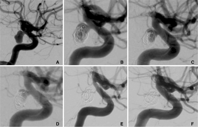
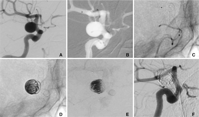
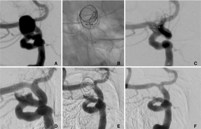
We identified 25 non-consecutive patients. The treatment was staged in 9 patients and in a single session 16 patients. The average age was 61±12.8 years (range 40-82). The average fundus height was 11±3.6 mm and average fundus width was 10.1±3.4 mm. In the staged cohort (n=9) at delayed angiography (mean 10 mths) 8 aneurysms (89%) showed complete exclusion (mRRC 1) and in one patient there was a parent vessel occlusion. In the simultaneous cohort delayed angiography (n=10, mean 8.1 months) demonstrated complete occlusion (mRRC 1) in 6 aneurysms (60%), 3 neck remnants (mRRC 2) (30%) and 1 patient (10%) showed persistent aneurysmal filling (mRRC 3a). There were 5 complications with permanent morbidity (mRS >2) in two patients. There were no mortalities.
The MED can be successfully used in combination with intraluminal FDS and in selected aneurysms this may represent an alternative to FDS and adjunctive coiling.
麦地那栓塞装置(MED)是一种新型囊内装置,早期结果令人期待。此前我们记录了单独使用该装置以及与包括血流导向支架(FDS)在内的其他装置联合使用的初步经验。我们试图确定MED + FDS策略治疗特定动脉瘤的效果。
我们对前瞻性收集的数据进行回顾性分析,以确定所有使用MED和腔内FDS治疗动脉瘤的患者。我们展示了技术成功率、早期和中期血管造影随访结果以及临床结局数据。
我们确定了25例非连续患者。9例患者采用分期治疗,16例患者采用单次治疗。平均年龄为61±12.8岁(范围40 - 82岁)。平均瘤底高度为11±3.6 mm,平均瘤底宽度为10.1±3.4 mm。在分期治疗组(n = 9)延迟血管造影(平均10个月)时,8个动脉瘤(89%)显示完全闭塞(改良Raymond分级1级),1例患者出现载瘤血管闭塞。在同期治疗组延迟血管造影(n = 10,平均8.1个月)时,6个动脉瘤(60%)显示完全闭塞(改良Raymond分级1级),3个瘤颈残留(改良Raymond分级2级)(30%),1例患者(10%)显示动脉瘤持续充盈(改良Raymond分级3a级)。有2例患者出现5例永久性致残并发症(改良Rankin量表评分>2分)。无死亡病例。
MED可成功与腔内FDS联合使用,对于特定动脉瘤,这可能是FDS和辅助弹簧圈栓塞的一种替代方法。