• 文献检索
  • 文档翻译
  • 深度研究
  • 学术资讯
  • Suppr Zotero 插件Zotero 插件
  • 邀请有礼
  • 套餐&价格
  • 历史记录
应用&插件
Suppr Zotero 插件Zotero 插件浏览器插件Mac 客户端Windows 客户端微信小程序
定价
高级版会员购买积分包购买API积分包
服务
文献检索文档翻译深度研究API 文档MCP 服务
关于我们
关于 Suppr公司介绍联系我们用户协议隐私条款
关注我们

Suppr 超能文献

核心技术专利:CN118964589B侵权必究
粤ICP备2023148730 号-1Suppr @ 2026

文献检索

告别复杂PubMed语法,用中文像聊天一样搜索,搜遍4000万医学文献。AI智能推荐,让科研检索更轻松。

立即免费搜索

文件翻译

保留排版,准确专业,支持PDF/Word/PPT等文件格式,支持 12+语言互译。

免费翻译文档

深度研究

AI帮你快速写综述,25分钟生成高质量综述,智能提取关键信息,辅助科研写作。

立即免费体验

Pin1 介导的组织因子基因表达、蛋白半衰期和促凝血活性增强的肽基脯氨酰顺反异构酶结构和细胞机制。

Structural and cellular mechanisms of peptidyl-prolyl isomerase Pin1-mediated enhancement of Tissue Factor gene expression, protein half-life, and pro-coagulant activity.

机构信息

Department of Medical Biochemistry, Amsterdam Cardiovascular Sciences, Academic Medical Center, University of Amsterdam, the Netherlands.

Department of Cell and Chemical Biology, Leiden University Medical Center, the Netherlands.

出版信息

Haematologica. 2018 Jun;103(6):1073-1082. doi: 10.3324/haematol.2017.183087. Epub 2018 Mar 15.

DOI:10.3324/haematol.2017.183087
PMID:29545340
原文链接:https://pmc.ncbi.nlm.nih.gov/articles/PMC6058786/
Abstract

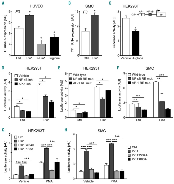
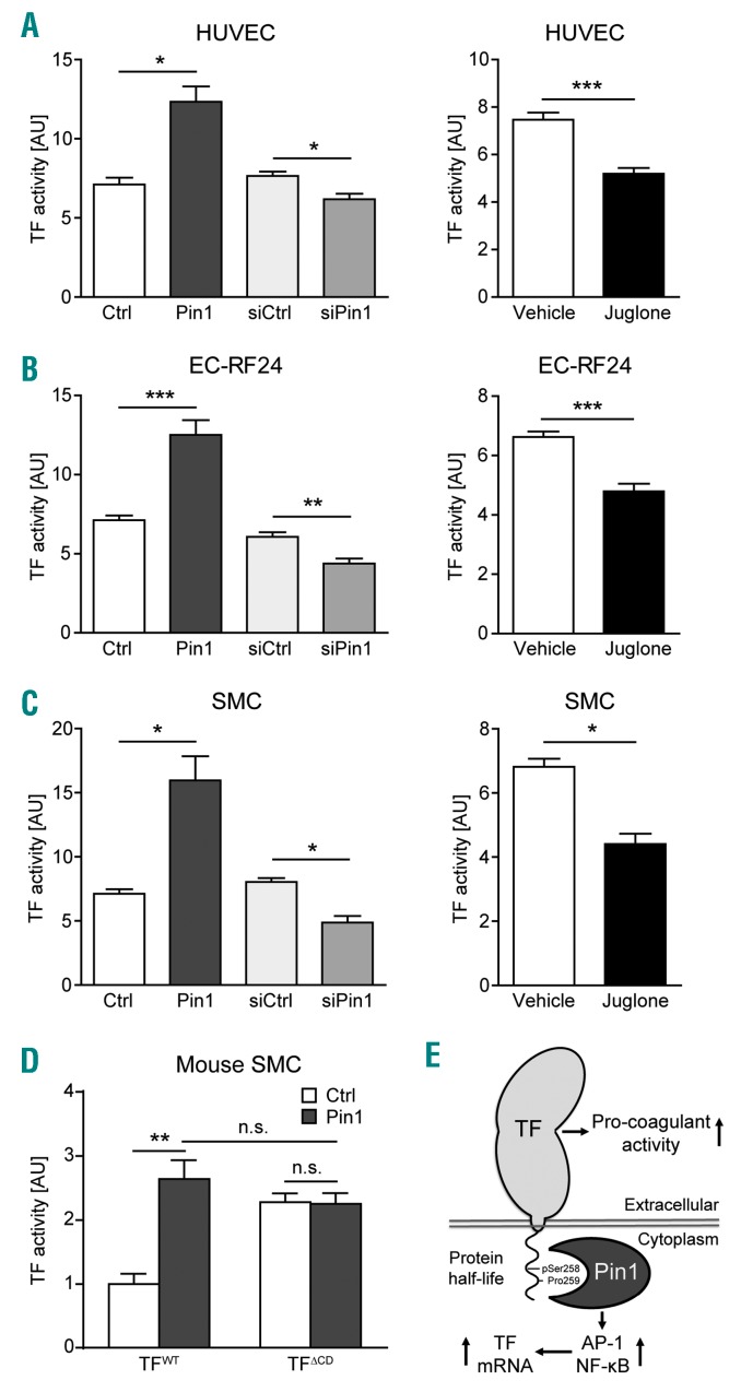
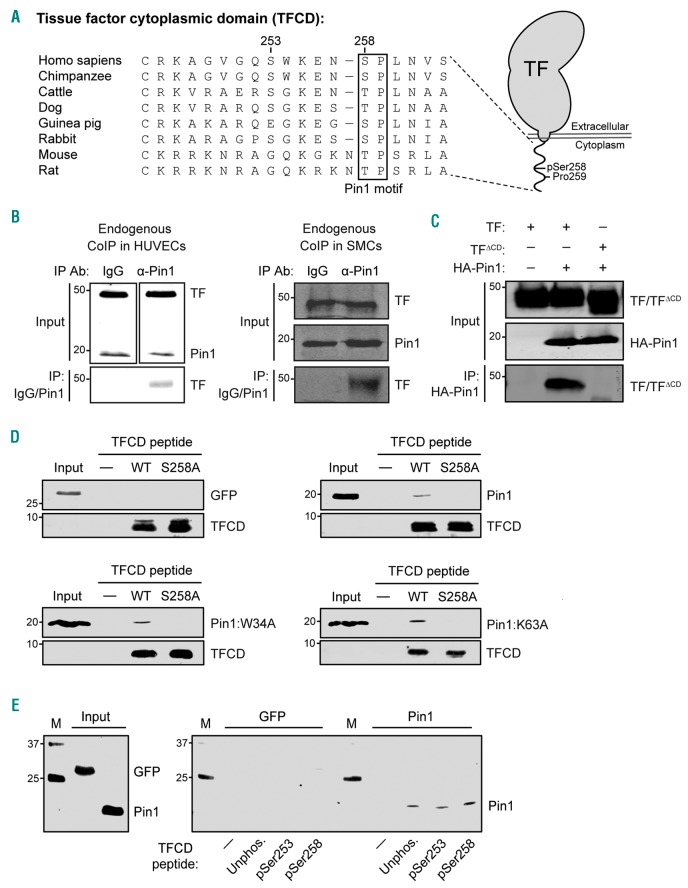
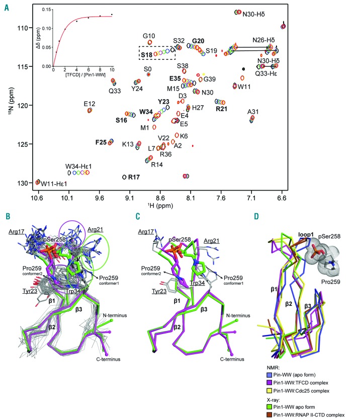
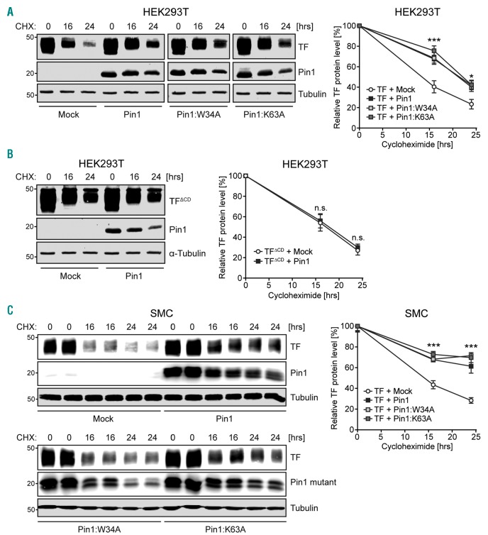
Tissue Factor is a cell-surface glycoprotein expressed in various cells of the vasculature and is the principal regulator of the blood coagulation cascade and hemostasis. Notably, aberrant expression of Tissue Factor is associated with cardiovascular pathologies such as atherosclerosis and thrombosis. Here, we sought to identify factors that regulate Tissue Factor gene expression and activity. Tissue Factor gene expression is regulated by various transcription factors, including activating protein-1 and nuclear factor-κ B. The peptidyl-prolyl isomerase Pin1 is known to modulate the activity of these two transcription factors, and we now show that Pin1 augments Tissue Factor gene expression in both vascular smooth muscle cells and activated endothelial cells activating protein-1 and nuclear factor-κ B signaling. Furthermore, the cytoplasmic domain of Tissue Factor contains a well-conserved phospho-Ser258-Pro259 amino-acid motif recognized by Pin1. Using co-immunoprecipitation and solution nuclear magnetic resonance spectroscopy, we show that the WW-domain of Pin1 directly binds the cytoplasmic domain of Tissue Factor. This interaction occurs the phospho-Ser258-Pro259 sequence in the Tissue Factor cytoplasmic domain and results in increased protein half-life and pro-coagulant activity. Taken together, our results establish Pin1 as an upstream regulator of Tissue Factor-mediated coagulation, thereby opening up new avenues for research into the use of specific Pin1 inhibitors for the treatment of diseases characterized by pathological coagulation, such as thrombosis and atherosclerosis.

摘要

组织因子是一种在血管的各种细胞中表达的细胞表面糖蛋白,是血液凝固级联和止血的主要调节剂。值得注意的是,组织因子的异常表达与心血管病理学有关,如动脉粥样硬化和血栓形成。在这里,我们试图确定调节组织因子基因表达和活性的因素。组织因子基因表达受多种转录因子调节,包括激活蛋白-1 和核因子-κB。肽基脯氨酰顺反异构酶 Pin1 已知调节这两种转录因子的活性,我们现在表明 Pin1 增强了血管平滑肌细胞和激活的内皮细胞中组织因子基因的表达,激活蛋白-1 和核因子-κB 信号。此外,组织因子的细胞质结构域包含一个保守的磷酸丝氨酸 258-脯氨酸 259 氨基酸基序,被 Pin1 识别。通过共免疫沉淀和溶液核磁共振波谱,我们表明 Pin1 的 WW 结构域直接与组织因子的细胞质结构域结合。这种相互作用发生在组织因子细胞质结构域的磷酸丝氨酸 258-脯氨酸 259 序列上,导致蛋白半衰期和促凝血活性增加。总之,我们的结果确立了 Pin1 作为组织因子介导的凝血的上游调节剂,从而为研究使用特定的 Pin1 抑制剂治疗以病理性凝血为特征的疾病(如血栓形成和动脉粥样硬化)开辟了新的途径。

https://cdn.ncbi.nlm.nih.gov/pmc/blobs/8c09/6058786/c8952701d396/1031073.fig5.jpg
https://cdn.ncbi.nlm.nih.gov/pmc/blobs/8c09/6058786/2b208c1382a2/1031073.fig1.jpg
https://cdn.ncbi.nlm.nih.gov/pmc/blobs/8c09/6058786/402fbbeeb9d0/1031073.fig2.jpg
https://cdn.ncbi.nlm.nih.gov/pmc/blobs/8c09/6058786/86155077bd37/1031073.fig3.jpg
https://cdn.ncbi.nlm.nih.gov/pmc/blobs/8c09/6058786/ec24adf84e16/1031073.fig4.jpg
https://cdn.ncbi.nlm.nih.gov/pmc/blobs/8c09/6058786/c8952701d396/1031073.fig5.jpg

相似文献

1
Structural and cellular mechanisms of peptidyl-prolyl isomerase Pin1-mediated enhancement of Tissue Factor gene expression, protein half-life, and pro-coagulant activity.Pin1 介导的组织因子基因表达、蛋白半衰期和促凝血活性增强的肽基脯氨酰顺反异构酶结构和细胞机制。
Haematologica. 2018 Jun;103(6):1073-1082. doi: 10.3324/haematol.2017.183087. Epub 2018 Mar 15.
2
Peptidyl-prolyl isomerase 1 (Pin1) preserves the phosphorylation state of tissue factor and prolongs its release within microvesicles.肽基脯氨酰顺反异构酶 1(Pin1)可保持组织因子的磷酸化状态,并延长其在微泡中的释放。
Biochim Biophys Acta Mol Cell Res. 2018 Jan;1865(1):12-24. doi: 10.1016/j.bbamcr.2017.09.016. Epub 2017 Sep 28.
3
The peptidyl prolyl isomerase, PIN1 induces angiogenesis through direct interaction with HIF-2α.肽基脯氨酰顺反异构酶 PIN1 通过与 HIF-2α 的直接相互作用诱导血管生成。
Biochem Biophys Res Commun. 2020 Dec 17;533(4):995-1003. doi: 10.1016/j.bbrc.2020.08.015. Epub 2020 Oct 2.
4
Peptidyl-prolyl isomerase Pin1 markedly enhances the oncogenic activity of the rel proteins in the nuclear factor-kappaB family.肽基脯氨酰顺反异构酶Pin1显著增强核因子-κB家族中Rel蛋白的致癌活性。
Cancer Res. 2009 Jun 1;69(11):4589-97. doi: 10.1158/0008-5472.CAN-08-4117. Epub 2009 May 19.
5
Peptidyl-prolyl isomerase Pin1 deficiency attenuates angiotensin II-induced abdominal aortic aneurysm formation in ApoE mice.肽基脯氨酰顺反异构酶 Pin1 缺乏可减轻载脂蛋白 E 小鼠血管紧张素Ⅱ诱导的腹主动脉瘤形成。
J Mol Cell Cardiol. 2018 Jan;114:334-344. doi: 10.1016/j.yjmcc.2017.12.006. Epub 2017 Dec 19.
6
Neighboring phosphoSer-Pro motifs in the undefined domain of IRAK1 impart bivalent advantage for Pin1 binding.在IRAK1未定义结构域中相邻的磷酸化丝氨酸-脯氨酸基序赋予Pin1结合的双价优势。
FEBS J. 2016 Dec;283(24):4528-4548. doi: 10.1111/febs.13943. Epub 2016 Nov 20.
7
Structural Analysis of the Pin1-CPEB1 interaction and its potential role in CPEB1 degradation.Pin1与CPEB1相互作用的结构分析及其在CPEB1降解中的潜在作用。
Sci Rep. 2015 Oct 12;5:14990. doi: 10.1038/srep14990.
8
Regulation of Pin1 peptidyl-prolyl cis/trans isomerase activity by its WW binding module on a multi-phosphorylated peptide of Tau protein.Pin1肽基脯氨酰顺/反异构酶活性通过其WW结合模块对Tau蛋白多磷酸化肽段的调控。
FEBS Lett. 2005 Aug 1;579(19):4159-64. doi: 10.1016/j.febslet.2005.06.048.
9
Negative Regulation of Peptidyl-Prolyl Isomerase Activity by Interdomain Contact in Human Pin1.人源Pin1中结构域间相互作用对肽基脯氨酰异构酶活性的负调控
Structure. 2015 Dec 1;23(12):2224-2233. doi: 10.1016/j.str.2015.08.019. Epub 2015 Oct 22.
10
Pin1 Modulates the Synaptic Content of NMDA Receptors via Prolyl-Isomerization of PSD-95.Pin1通过PSD-95的脯氨酰异构化调节NMDA受体的突触含量。
J Neurosci. 2016 May 18;36(20):5437-47. doi: 10.1523/JNEUROSCI.3124-15.2016.

引用本文的文献

1
Pin1-Catalyzed Conformation Changes Regulate Protein Ubiquitination and Degradation.Pin1 催化的构象变化调节蛋白质泛素化和降解。
Cells. 2024 Apr 23;13(9):731. doi: 10.3390/cells13090731.
2
Regulation of tissue factor activity by interaction with the first PDZ domain of MAGI1.通过与MAGI1的第一个PDZ结构域相互作用对组织因子活性的调节
Thromb J. 2024 Jan 17;22(1):12. doi: 10.1186/s12959-023-00580-6.
3
The Tissue Factor Pathway in Cancer: Overview and Role of Heparan Sulfate Proteoglycans.癌症中的组织因子途径:硫酸乙酰肝素蛋白聚糖的概述及作用

本文引用的文献

1
Identification of the integrin-binding site on coagulation factor VIIa required for proangiogenic PAR2 signaling.鉴定凝血因子 VIIa 上整合素结合位点对于 PAR2 信号的促血管生成作用是必需的。
Blood. 2018 Feb 8;131(6):674-685. doi: 10.1182/blood-2017-02-768218. Epub 2017 Dec 15.
2
Peptidyl-prolyl isomerase 1 (Pin1) preserves the phosphorylation state of tissue factor and prolongs its release within microvesicles.肽基脯氨酰顺反异构酶 1(Pin1)可保持组织因子的磷酸化状态,并延长其在微泡中的释放。
Biochim Biophys Acta Mol Cell Res. 2018 Jan;1865(1):12-24. doi: 10.1016/j.bbamcr.2017.09.016. Epub 2017 Sep 28.
3
Oligoubiquitination of tissue factor on Lys255 promotes Ser253-dephosphorylation and terminates TF release.
Cancers (Basel). 2023 Feb 28;15(5):1524. doi: 10.3390/cancers15051524.
4
Tissue factor as a new target for tumor therapy-killing two birds with one stone: a narrative review.组织因子作为肿瘤治疗的新靶点——一石二鸟:一篇叙述性综述
Ann Transl Med. 2022 Nov;10(22):1250. doi: 10.21037/atm-22-5067.
5
Tissue factor in cancer-associated thromboembolism: possible mechanisms and clinical applications.癌症相关血栓栓塞中的组织因子:可能的机制和临床应用。
Br J Cancer. 2022 Dec;127(12):2099-2107. doi: 10.1038/s41416-022-01968-3. Epub 2022 Sep 12.
6
Pin1 as Molecular Switch in Vascular Endothelium: Notes on Its Putative Role in Age-Associated Vascular Diseases.Pin1 作为血管内皮中的分子开关:其在与年龄相关的血管疾病中潜在作用的注释。
Cells. 2021 Nov 24;10(12):3287. doi: 10.3390/cells10123287.
7
Inhibition of the prolyl isomerase Pin1 improves endothelial function and attenuates vascular remodelling in pulmonary hypertension by inhibiting TGF-β signalling.脯氨酰异构酶 Pin1 的抑制作用通过抑制 TGF-β 信号通路改善肺动脉高压中的内皮功能并减弱血管重构。
Angiogenesis. 2022 Feb;25(1):99-112. doi: 10.1007/s10456-021-09812-7. Epub 2021 Aug 11.
8
Endothelial Dysfunction in Pulmonary Hypertension: Cause or Consequence?肺动脉高压中的内皮功能障碍:原因还是结果?
Biomedicines. 2021 Jan 9;9(1):57. doi: 10.3390/biomedicines9010057.
9
Murine tissue factor disulfide mutation causes a bleeding phenotype with sex specific organ pathology and lethality.鼠组织因子二硫键突变导致具有性别特异性器官病理学和致死性的出血表型。
Haematologica. 2020 Oct 1;105(10):2484-2495. doi: 10.3324/haematol.2019.218818.
10
Alternative activation of human macrophages enhances tissue factor expression and production of extracellular vesicles.人巨噬细胞的替代激活增强了组织因子的表达和细胞外囊泡的产生。
Haematologica. 2021 Feb 1;106(2):454-463. doi: 10.3324/haematol.2019.220210.
组织因子赖氨酸255位点的寡聚泛素化促进丝氨酸253去磷酸化并终止组织因子释放。
Biochim Biophys Acta. 2016 Nov;1863(11):2846-2857. doi: 10.1016/j.bbamcr.2016.09.005. Epub 2016 Sep 4.
4
Pre-Anchoring of Pin1 to Unphosphorylated c-Myc in a Fuzzy Complex Regulates c-Myc Activity.在模糊复合物中Pin1与未磷酸化的c-Myc的预锚定调节c-Myc活性。
Structure. 2015 Dec 1;23(12):2267-2279. doi: 10.1016/j.str.2015.10.010. Epub 2015 Nov 19.
5
Structural Analysis of the Pin1-CPEB1 interaction and its potential role in CPEB1 degradation.Pin1与CPEB1相互作用的结构分析及其在CPEB1降解中的潜在作用。
Sci Rep. 2015 Oct 12;5:14990. doi: 10.1038/srep14990.
6
Non-catalytic participation of the Pin1 peptidyl-prolyl isomerase domain in target binding.Pin1 肽基脯氨酰顺反异构酶结构域的非催化靶标结合。
Front Physiol. 2013 Feb 13;4:18. doi: 10.3389/fphys.2013.00018. eCollection 2013.
7
New fundamentals in hemostasis.止血的新基础。
Physiol Rev. 2013 Jan;93(1):327-58. doi: 10.1152/physrev.00016.2011.
8
p38α phosphorylates serine 258 within the cytoplasmic domain of tissue factor and prevents its incorporation into cell-derived microparticles.p38α使组织因子胞质结构域内的丝氨酸258磷酸化,并阻止其掺入细胞衍生的微粒中。
Biochim Biophys Acta. 2013 Mar;1833(3):613-21. doi: 10.1016/j.bbamcr.2012.11.010. Epub 2012 Nov 26.
9
An unusual two-step control of CPEB destruction by Pin1.Pin1 通过一种不寻常的两步控制 CPEB 的破坏。
Mol Cell Biol. 2013 Jan;33(1):48-58. doi: 10.1128/MCB.00904-12. Epub 2012 Oct 22.
10
Murine tissue factor coagulant activity is critically dependent on the presence of an intact allosteric disulfide.鼠组织因子凝血活性严重依赖于完整的变构二硫键的存在。
Haematologica. 2013 Jan;98(1):153-8. doi: 10.3324/haematol.2012.069997. Epub 2012 Jul 16.