Tanisaka Yuki, Ryozawa Shomei, Kobayashi Masanori, Harada Maiko, Kobatake Tsutomu, Omiya Kumiko, Iwano Hirotoshi, Arai Shin, Nonaka Kouichi, Mashimo Yumi
Department of Gastroenterology, Saitama Medical University International Medical Center, Hidaka, Saitama 350-1298, Japan.
Oncol Lett. 2018 Apr;15(4):4759-4766. doi: 10.3892/ol.2018.7939. Epub 2018 Feb 2.
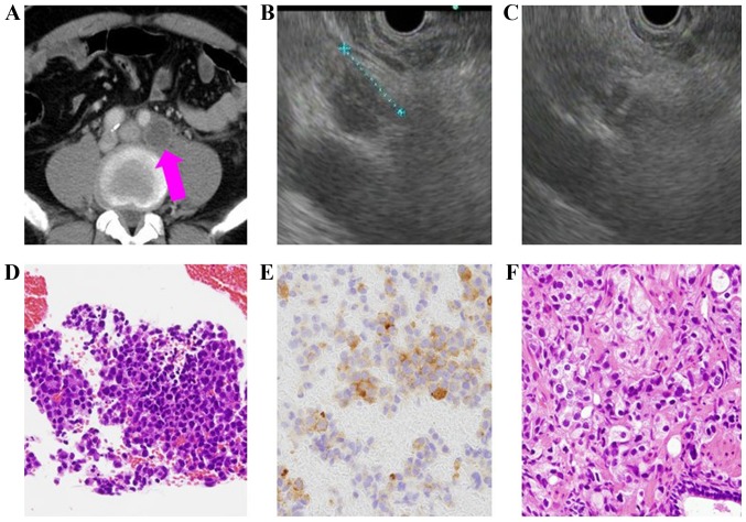
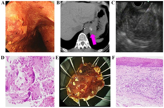
Lymphadenopathy may be difficult to diagnose using imaging results alone. Endoscopic ultrasound-guided fine needle aspiration (EUS-FNA) may help to diagnose and determine the appropriate management of lymphadenopathy. EUS-FNA has been used as a safe and less invasive method for obtaining pathologic specimens from extraluminal lesions using endoscopic ultrasound. The present study evaluated the usefulness of EUS-FNA for lymphadenopathy. Between July 2013 and December 2016, 72 patients undergoing EUS-FNA for lymphadenopathy that could not be diagnosed solely using imaging were included. The present study evaluated the sensitivity, specificity, positive and negative predictive value, overall accuracy, helpfulness in determining the management of lymphadenopathy and EUS-FNA-associated complications. Of the 72 included patients, 8 were diagnosed with benign (inflammatory or reactive) lymphadenopathy. The diagnostic sensitivity, specificity, positive and negative predictive value, and overall accuracy were 95.3, 100, 100, 72.7 and 95.8%, respectively. While EUS-FNA of metastatic nodes identified the origin in the majority of cases, the procedure resulted in a different histopathological diagnosis from the previous image-based diagnosis in 9 patients. Consequently, 2 patients with testicular cancer were administered bleomycin, etoposide, and cisplatin. An individual with GIST was administered imatinib, and a patient with prostate cancer was administered degarelix (antihormon drug). A total of 5 other patients received palliative medicine due to the change in diagnosis. EUS-FNA also helped determine the appropriate cancer management plan in other patients; specifically, based on the cytology of the metastatic lymph node, EUS-FNA helped determine the cancer stage, and to identify recurrence or the primary cancer from which tissue could not be collected. No EUS-FNA-associated symptoms were reported. To conclude, the present study suggested that EUS-FNA of suspected metastatic lymph nodes appears safe and useful for cancer staging and diagnosing recurrence. It may also useful for diagnosing patients whose collection of samples from the original cancer appeared impractical. EUS-FNA for lymphadenopathy that may not be diagnosed with imaging alone may assist in diagnosis and help to determine the appropriate management strategy.
仅依靠影像学结果可能难以诊断淋巴结病。内镜超声引导下细针穿刺抽吸术(EUS-FNA)有助于诊断淋巴结病并确定适当的治疗方法。EUS-FNA已被用作一种安全且侵入性较小的方法,通过内镜超声从腔外病变获取病理标本。本研究评估了EUS-FNA对淋巴结病的实用性。在2013年7月至2016年12月期间,纳入了72例因仅靠影像学无法诊断而接受EUS-FNA检查的淋巴结病患者。本研究评估了其敏感性、特异性、阳性和阴性预测值、总体准确性、在确定淋巴结病治疗方法方面的帮助以及与EUS-FNA相关的并发症。在纳入的72例患者中,8例被诊断为良性(炎症性或反应性)淋巴结病。诊断敏感性、特异性、阳性和阴性预测值以及总体准确性分别为95.3%、100%、100%、72.7%和95.8%。虽然转移性淋巴结的EUS-FNA在大多数情况下确定了起源,但该操作导致9例患者的组织病理学诊断与先前基于图像的诊断不同。因此,2例睾丸癌患者接受了博来霉素、依托泊苷和顺铂治疗。1例胃肠道间质瘤患者接受了伊马替尼治疗,1例前列腺癌患者接受了地加瑞克(抗激素药物)治疗。另外共有5例患者因诊断改变而接受了姑息治疗。EUS-FNA还帮助其他患者确定了适当的癌症治疗方案;具体而言,基于转移性淋巴结的细胞学检查,EUS-FNA有助于确定癌症分期,并从无法获取组织的部位识别复发或原发性癌症。未报告与EUS-FNA相关的症状。总之,本研究表明,对疑似转移性淋巴结进行EUS-FNA似乎对癌症分期和诊断复发是安全且有用的。它对于诊断那些从原发性癌症获取样本似乎不切实际的患者也可能有用。对仅靠影像学无法诊断的淋巴结病进行EUS-FNA可能有助于诊断并帮助确定适当的治疗策略。