Department of Oral and Maxillofacial Radiology, Aichi-Gakuin University School of Dentistry, Nagoya, Japan.
Department of Pharmacology, Aichi-Gakuin University School of Dentistry, Nagoya, Japan.
Int J Oral Sci. 2018 Mar 15;10(2):8. doi: 10.1038/s41368-017-0001-y.
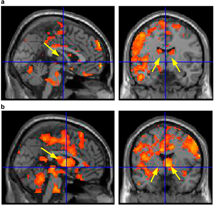
An animal experiment clarified that insertion of an orthodontic apparatus activated the trigeminal neurons of the medulla oblongata. Orthodontic tooth movement is known to be associated with the sympathetic nervous system and controlled by the nucleus of the hypothalamus. However, the transmission of both has not been demonstrated in humans. The purpose of this study were to examine the activated cerebral areas using brain functional magnetic resonance imaging (MRI), when orthodontic tooth separators were inserted, and to confirm the possibility of the transmission route from the medulla oblongata to the hypothalamus.
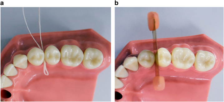
Two types of alternative orthodontic tooth separators (brass contact gauge and floss) were inserted into the right upper premolars of 10 healthy volunteers. Brain functional T2*-weighted images and anatomical T1-weighted images were taken.
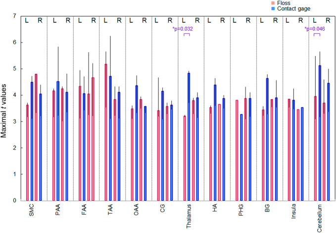
The blood oxygenation level dependent (BOLD) signals following insertion of a brass contact gauge and floss significantly increased in the somatosensory association cortex and hypothalamic area.
Our findings suggest the possibility of a transmission route from the medulla oblongata to the hypothalamus.
动物实验阐明,正畸装置的插入会激活延髓的三叉神经神经元。已知正畸牙齿移动与交感神经系统有关,并受下丘脑核控制。然而,这两者在人体中的传递尚未得到证实。本研究旨在使用脑功能磁共振成像(fMRI)检查插入正畸牙分离器时被激活的大脑区域,并确认从延髓到下丘脑的可能传递途径。
将两种不同类型的正畸牙分离器(黄铜接触规和牙线)插入 10 名健康志愿者的右上颌前磨牙中。采集脑功能 T2*-加权图像和解剖 T1-加权图像。
插入黄铜接触规和牙线后,感觉联合皮层和下丘脑区域的血氧水平依赖(BOLD)信号显著增加。
我们的发现表明,从延髓到下丘脑可能存在一种传递途径。