Shi Jing, Zhang Jialing, Yin Meng, Wang Qian, Du Jun
Imaging Diagnosis Center, Shanghai Children's Medical Center, Shanghai Jiao Tong University School of Medicine, Shanghai 200127, P.R. China.
Department of Cardiothoracic Surgery, Shanghai Children's Medical Center, Shanghai Jiao Tong University School of Medicine, Shanghai 200127, P.R. China.
Exp Ther Med. 2018 Apr;15(4):3899-3907. doi: 10.3892/etm.2018.5895. Epub 2018 Feb 26.
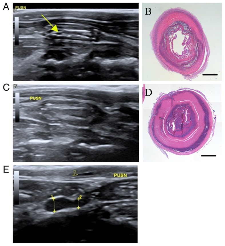
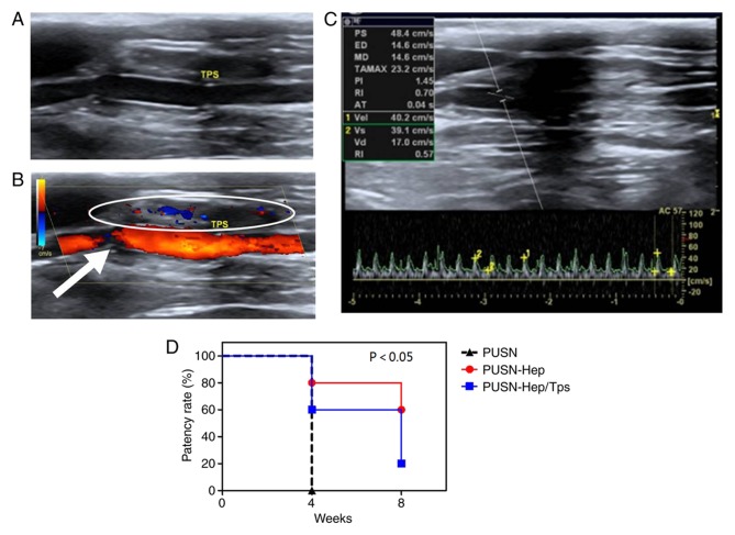
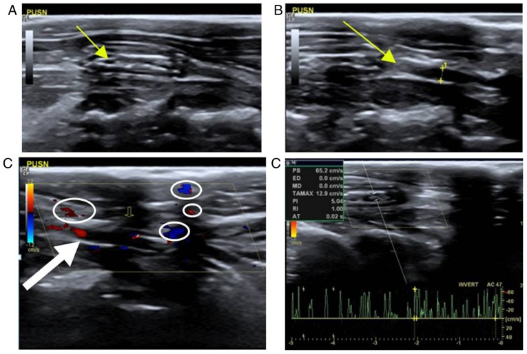
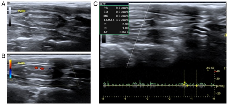
There is a large clinical requirement for novel vascular grafts; however, the development of novel vascular grafts has not been extremely successful to date. The most successful method for the continuous evaluation of vascular grafts remains unclear. Therefore, an optimal successive, non-invasive imaging modality is necessary for the study of vascular transplantation. In the present study, a common rabbit model of carotid artery defect was utilized. The patency and hemodynamic characteristics of implanted grafts was examined following surgery by color Doppler ultrasound in three modes, including B-mode, color flow map and pulse-Doppler examination. The results revealed that ultrasound had sufficient spatial resolution to generate clear images of the carotid artery of rabbits with or without the implanted grafts. Color Doppler ultrasound may be applied to evaluate and differentiate the patent, stenosis and occlusion of carotid arteries in rabbits with different vascular grafts implanted. Furthermore, color Doppler ultrasound is an optimal imaging modality for continuous evaluation . It is also possible for some quantitative analyses to be performed, including measuring the diameter of vascular lumens and the flow velocity of the region of interest. The present study suggests vascular ultrasound as the optimum choice for continuous surveillance of vascular prostheses , which may provide valuable information about the grafts in order to greatly shorten the experimental period.
对新型血管移植物有很大的临床需求;然而,迄今为止新型血管移植物的研发尚未取得巨大成功。目前仍不清楚对血管移植物进行持续评估的最成功方法。因此,对于血管移植研究而言,一种最佳的连续、非侵入性成像方式是必要的。在本研究中,使用了常见的兔颈动脉缺损模型。术后通过彩色多普勒超声的三种模式,包括B超模式、彩色血流图和脉冲多普勒检查,来检测植入移植物的通畅情况和血流动力学特征。结果显示,超声具有足够的空间分辨率,能够清晰显示植入或未植入移植物的兔颈动脉图像。彩色多普勒超声可用于评估和区分植入不同血管移植物的兔颈动脉的通畅、狭窄和闭塞情况。此外,彩色多普勒超声是进行连续评估的最佳成像方式。还可以进行一些定量分析,包括测量血管腔直径和感兴趣区域的血流速度。本研究表明,血管超声是对血管假体进行连续监测的最佳选择,其可为移植物提供有价值的信息,从而大幅缩短实验周期。