Department of Ophthalmology, Second People's Hospital of Yunnan Province, Kunming, Yunan 650021, P.R. China.
Department of Ophthalmology, Yan'an Hospital, Kunming, Yunan 650051, P.R. China.
Mol Med Rep. 2018 May;17(5):7177-7183. doi: 10.3892/mmr.2018.8738. Epub 2018 Mar 14.
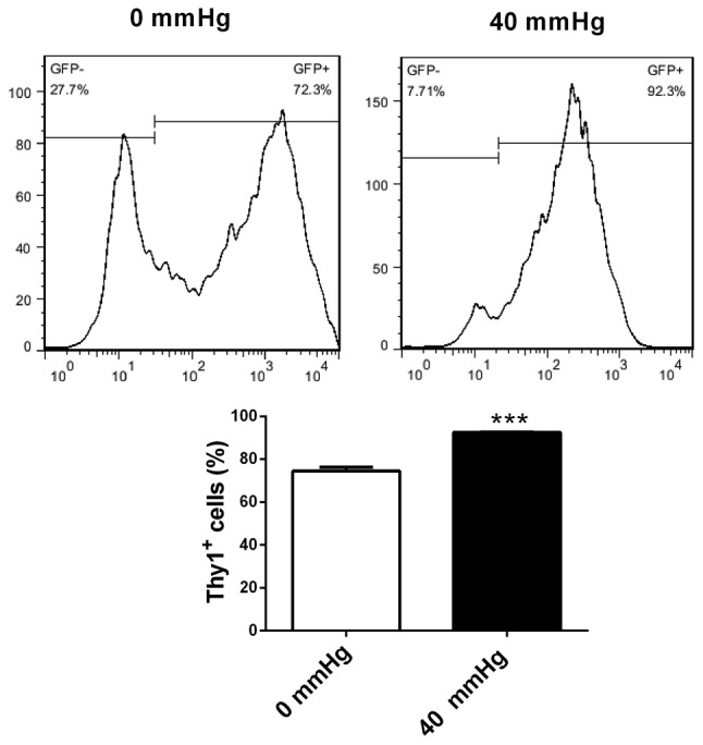
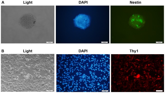
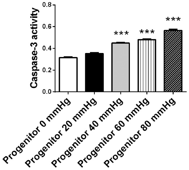
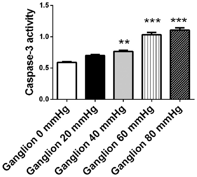
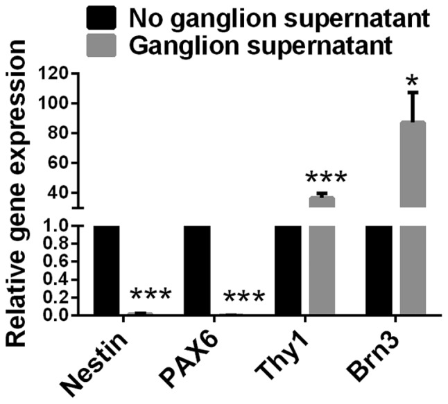
Loss of retinal ganglion cells is implicated in glaucoma and high intraocular pressure. Factors that affect the differentiation of retinal progenitor cells into retinal ganglion cells remain unclear. The present study aimed to investigate the effects of retinal ganglion cell‑conditioned medium on gene expression and differentiation in retinal progenitor cells, and the effects of surrounding pressure on the survival and differentiation of retinal progenitor cells. Retinal progenitor cells and retinal ganglion cells were isolated from rats. Immunofluorescence staining of Nestin and Thy1 was performed to identify rat retinal progenitor cells and retinal ganglion cells, respectively. Retinal progenitor cells and ganglion cells were cultured for 48 h under surrounding pressure of 0, 20, 40, 60 and 80 mmHg. Cellular apoptosis was detected using a caspase‑3 assay kit. In addition, the culture supernatant of rat retinal ganglion cells was collected. Retinal progenitor cells were cultured in the presence or absence of retinal ganglion‑conditioned medium for 72 h under normal pressure. Gene expression of Nestin, paired box protein 6 (PAX6), Thy1 and brain‑specific homeobox/POU domain protein 3 (Brn‑3) in retinal progenitor cells was detected by reverse transcription‑quantitative polymerase chain reaction. Retinal progenitor cells were cultured in retinal ganglion‑conditioned medium for 72 h under surrounding pressure of 0 and 40 mmHg, respectively, and flow cytometry was utilized to evaluate the effects of pressure on the differentiation of retinal progenitor cells into retinal ganglion cells. The results demonstrated that isolated retinal progenitor cells were Nestin‑positive and retinal ganglion cells were Thy1‑positive, suggesting successful isolation. The activity of caspase‑3 increased in retinal progenitor cells and retinal ganglion cells in a pressure‑dependent manner. When the surrounding pressure reached 40, 60 and 80 mmHg, the activity of caspase‑3 in retinal progenitor cells and ganglion cells increased significantly compared with cells that were not under pressure. Compared with retinal progenitor cells cultured without ganglion‑conditioned medium, those cultured with ganglion‑conditioned medium had significantly decreased expression levels of Nestin and PAX6, and increased expression levels of Thy1 and Brn3. Compared with 0 mmHg pressure, retinal progenitor cells cultured in ganglion‑conditioned medium under 40 mmHg pressure had increased percentages of Thy1‑positive cells. In conclusion, the apoptosis of rat retinal progenitor cells and retinal ganglion cells was pressure‑dependent. Retinal ganglion cell‑conditioned medium increased the differentiation of retinal progenitor cells into retinal ganglion‑like cells, and the differentiation increased as surrounding pressure increased. Current study provides insights that may contribute to the efforts of developing a treatment for glaucoma.
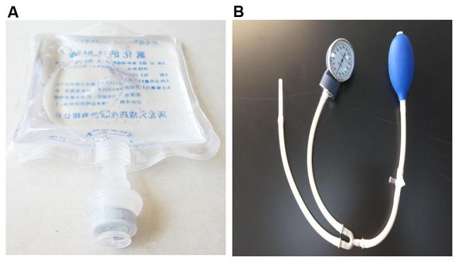
视网膜神经节细胞的丢失与青光眼和眼内压升高有关。影响视网膜祖细胞分化为视网膜神经节细胞的因素尚不清楚。本研究旨在探讨视网膜神经节细胞条件培养基对视网膜祖细胞基因表达和分化的影响,以及周围压力对视网膜祖细胞存活和分化的影响。从大鼠中分离出视网膜祖细胞和视网膜神经节细胞。分别通过巢蛋白和 Thy1 的免疫荧光染色鉴定大鼠视网膜祖细胞和视网膜神经节细胞。在 0、20、40、60 和 80mmHg 的周围压力下培养视网膜祖细胞和神经节细胞 48h。使用 caspase-3 测定试剂盒检测细胞凋亡。此外,收集大鼠视网膜神经节细胞的培养上清液。在正常压力下,将视网膜祖细胞在有无视网膜神经节条件培养基的情况下培养 72h。通过逆转录定量聚合酶链反应检测视网膜祖细胞中巢蛋白、配对盒蛋白 6 (PAX6)、Thy1 和脑特异性同源盒/POU 结构域蛋白 3 (Brn-3)的基因表达。在 0 和 40mmHg 的周围压力下,将视网膜祖细胞分别在视网膜神经节条件培养基中培养 72h,然后使用流式细胞术评估压力对视网膜祖细胞分化为视网膜神经节细胞的影响。结果表明,分离的视网膜祖细胞巢蛋白阳性,视网膜神经节细胞 Thy1 阳性,提示成功分离。caspase-3 的活性随压力呈依赖性增加。当周围压力达到 40、60 和 80mmHg 时,视网膜祖细胞和神经节细胞中的 caspase-3 活性明显高于无压力的细胞。与未培养在神经节条件培养基中的视网膜祖细胞相比,培养在神经节条件培养基中的视网膜祖细胞巢蛋白和 PAX6 的表达水平显著降低,Thy1 和 Brn3 的表达水平显著升高。与 0mmHg 压力相比,在神经节条件培养基中培养的视网膜祖细胞在 40mmHg 压力下 Thy1 阳性细胞的百分比增加。综上所述,大鼠视网膜祖细胞和神经节细胞的凋亡与压力有关。视网膜神经节细胞条件培养基增加了视网膜祖细胞向视网膜神经节样细胞的分化,并且随着周围压力的增加,分化增加。本研究为开发青光眼治疗方法提供了新的思路。