Department of Radiation Oncology, University Hospital of Heidelberg, Im Neuenheimer Feld 400, 69120, Heidelberg, Germany.
Heidelberg Institute for Radiation Oncology (HIRO), Im Neuenheimer Feld 400, 69120, Heidelberg, Germany.
Radiat Oncol. 2018 Mar 27;13(1):54. doi: 10.1186/s13014-018-1002-5.
Meningiomas of the skull base account for 25-30% of all meningiomas. Due to the complex structure of the cranial base and its close proximity to critical structures, surgery is often associated with substantial morbidity. Treatment options include observation, aggressive surgical intervention, stereotactic or conventional radiotherapy. In this analysis we evaluate the outcome of 110 patients with meningiomas of the skull base treated with particle therapy. It was performed within the framework of the "clinical research group heavy ion therapy" and supported by the German Research Council (DFG, KFO 214).
Between May 2010 and November 2014, 110 Patients with skull base meningioma were treated with particle radiotherapy at the Heidelberg Ion Therapy Center (HIT). Primary localizations included the sphenoid wing (n = 42), petroclival region (n = 23), cavernous sinus (n = 4), sella (n = 10) and olfactory nerve (n = 4). Sixty meningiomas were benign (WHO °I); whereas 8 were high-risk (WHO °II (n = 7) and °III (n = 1)). In 42 cases histology was not examined, since no surgery was performed. Proton (n = 104) or carbon ion (n = 6) radiotherapy was applied at Heidelberg Ion Therapy Center (HIT) using raster-scanning technique for active beam delivery. Fifty one patients (46.4%) received radiotherapy due to tumor progression, 17 (15.5%) after surgical resection and 42 (38.2%) as primary treatment.
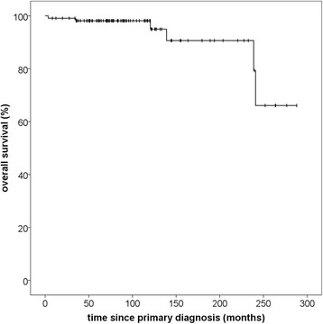
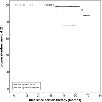
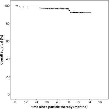
Median follow-up in this analysis was 46,8 months (95% CI 39,9-53,7; Q1-Q3 34,3-61,7). Particle radiotherapy could be performed safely without toxicity-related interruptions. No grade IV or V toxicities according to CTCAE v4.0 were observed. Particle RT offered excellent overall local control rates with 100% progression-free survival (PFS) after 36 months and 96.6% after 60 months. Median PFS was not reached due to the small number of events. Histology significantly impacted PFS with superior PFS after 5 years for low-risk tumors (96.6% vs. 75.0%, p = 0,02). Overall survival was 96.2% after 60 months and 92.0% after 72 months from therapy. Of six documented deaths, five were definitely not and the sixth probably not meningioma-related.
Particle radiotherapy is an excellent treatment option for patients with meningiomas of the skull base and can lead to long-term tumor control with minimal side effects. Other prospective studies with longer follow-up will be necessary to further confirm the role of particle radiotherapy in skull base meningioma.
颅底脑膜瘤占所有脑膜瘤的 25-30%。由于颅底结构复杂,且紧邻关键结构,手术常伴有较高的发病率。治疗选择包括观察、积极手术干预、立体定向或常规放疗。在这项分析中,我们评估了 110 例接受粒子治疗的颅底脑膜瘤患者的结果。该分析是在“重离子治疗临床研究组”的框架内进行的,并得到了德国研究基金会(DFG,KFO 214)的支持。
2010 年 5 月至 2014 年 11 月,110 例颅底脑膜瘤患者在海德堡离子治疗中心(HIT)接受粒子放疗。主要原发部位包括蝶骨翼(n=42)、岩斜区(n=23)、海绵窦(n=4)、蝶鞍(n=10)和嗅神经(n=4)。60 例脑膜瘤为良性(WHO °I);8 例为高风险(WHO °II(n=7)和 °III(n=1))。42 例因未行手术而未行组织学检查。质子(n=104)或碳离子(n=6)放疗在海德堡离子治疗中心(HIT)使用栅格扫描技术进行主动束输送。51 例(46.4%)因肿瘤进展接受放疗,17 例(15.5%)因手术切除后接受放疗,42 例(38.2%)作为初始治疗。
在这项分析中,中位随访时间为 46.8 个月(95%CI 39.9-53.7;Q1-Q3 34.3-61.7)。粒子放疗可安全进行,无毒性相关中断。未观察到 CTCAE v4.0 分级 IV 或 V 级毒性。粒子 RT 提供了出色的整体局部控制率,36 个月时无进展生存率(PFS)为 100%,60 个月时为 96.6%。由于事件数量较少,中位 PFS 未达到。组织学显著影响 PFS,低危肿瘤 5 年 PFS 更高(96.6% vs. 75.0%,p=0.02)。60 个月时的总生存率为 96.2%,72 个月时为 92.0%。在 6 例记录的死亡中,5 例肯定不是,第 6 例可能不是脑膜瘤相关。
粒子放疗是颅底脑膜瘤患者的一种极好的治疗选择,可以通过最小的副作用实现长期肿瘤控制。其他具有更长随访时间的前瞻性研究将有必要进一步证实粒子放疗在颅底脑膜瘤中的作用。