Sun Qian, Chen Guan-Qun, Wang Xi-Bin, Yu Ying, Hu Yu-Chuan, Yan Lin-Feng, Zhang Xin, Yang Yang, Zhang Jin, Liu Bin, Wang Cong-Cong, Ma Yi, Wang Wen, Han Ying, Cui Guang-Bin
Department of Radiology & Functional and Molecular Imaging, Key Lab of Shaanxi Province, Tangdu Hospital, The Military Medical University of PLA Airforce (Fourth Military Medical University), Xi'an, China.
Department of Neurology, XuanWu Hospital, Capital Medical University, Beijing, China.
Front Neuroanat. 2018 Mar 20;12:21. doi: 10.3389/fnana.2018.00021. eCollection 2018.
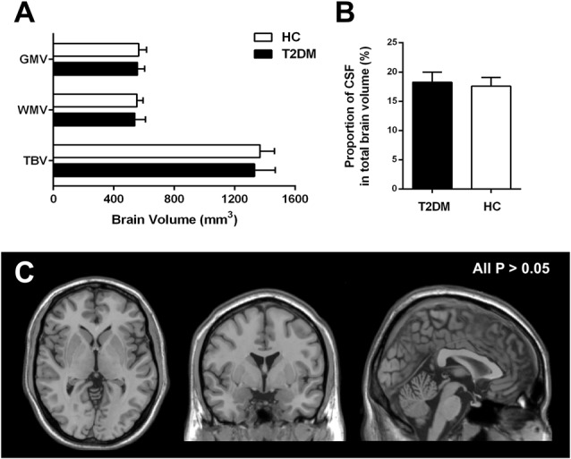
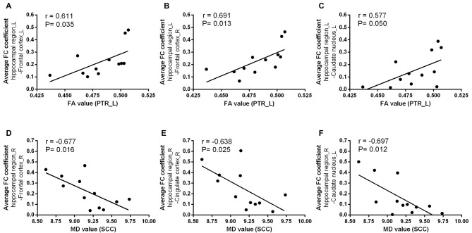
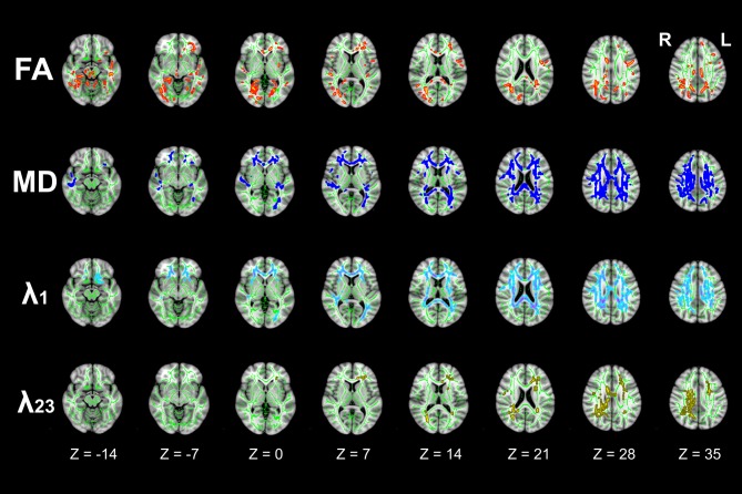
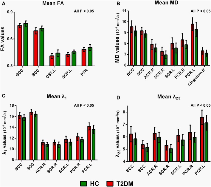
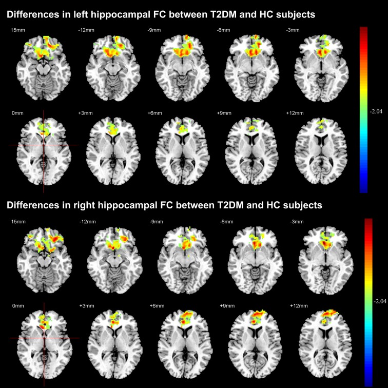
: To investigate the white matter (WM) integrity and hippocampal functional connectivity (FC) in type 2 diabetes mellitus (T2DM) patients without mild cognitive impairment (MCI) by using diffusion tensor imaging (DTI) and resting-state functional magnetic resonance imaging (rs-fMRI), respectively. : Twelve T2DM patients without MCI and 24 age, sex and education matched healthy controls (HC) were recruited. DTI and rs-fMRI data were subsequently acquired on a 3.0T MR scanner. Tract-based spatial statistics (TBSS) combining region of interests (ROIs) analysis was used to investigate the alterations of DTI metrics (fractional anisotropy (FA), mean diffusivity (MD), λ and λ) and FC measurement was performed to calculate hippocampal FC with other brain regions. Cognitive function was evaluated by using Mini-Mental State Examination (MMSE) and Montreal Cognitive Assessment (MoCA). Brain volumes were also evaluated among these participants. : There were no difference of MMSE and MoCA scores between two groups. Neither whole brain nor regional brain volume decrease was revealed in T2DM patients without MCI. DTI analysis revealed extensive WM disruptions, especially in the body of corpus callosum (CC). Significant decreases of hippocampal FC with certain brain structures were revealed, especially with the bilateral frontal cortex. Furthermore, the decreased FA in left posterior thalamic radiation (PTR) and increased MD in the splenium of CC were closely related with the decreased hippocampal FC to caudate nucleus and frontal cortex. : T2DM patients without MCI showed extensive WM disruptions and abnormal hippocampal FC. Moreover, the WM disruptions and abnormal hippocampal FC were closely associated. -T2DM patients without MCI demonstrated no obvious brain volume decrease.-Extensive white matter disruptions, especially within the body of corpus callosum, were revealed with DTI analysis among the T2DM patients.-Despite no MCI in T2DM patients, decreased functional connectivity between hippocampal region and some critical brain regions were detected.-The alterations in hippocampal functional connectivity were closely associated with those of the white matter structures in T2DM patients. This trial was registered to ClinicalTrials.gov (NCT02420470, https://www.clinicaltrials.gov/).
分别采用扩散张量成像(DTI)和静息态功能磁共振成像(rs-fMRI),研究无轻度认知障碍(MCI)的2型糖尿病(T2DM)患者的白质(WM)完整性和海马功能连接性(FC)。
招募了12名无MCI的T2DM患者和24名年龄、性别及教育程度相匹配的健康对照者(HC)。随后在3.0T磁共振扫描仪上采集DTI和rs-fMRI数据。采用基于纤维束的空间统计学(TBSS)结合感兴趣区(ROI)分析来研究DTI指标(分数各向异性(FA)、平均扩散率(MD)、λ1和λ2)的改变,并进行FC测量以计算海马与其他脑区的FC。使用简易精神状态检查表(MMSE)和蒙特利尔认知评估量表(MoCA)评估认知功能。还对这些参与者的脑容量进行了评估。
两组之间的MMSE和MoCA评分没有差异。无MCI的T2DM患者未发现全脑或局部脑容量减少。DTI分析显示广泛的白质破坏,尤其是在胼胝体(CC)体部。海马与某些脑结构的FC显著降低,尤其是与双侧额叶皮质。此外,左侧丘脑后辐射(PTR)的FA降低和CC压部的MD增加与海马与尾状核和额叶皮质的FC降低密切相关。
无MCI的T2DM患者表现出广泛的白质破坏和异常的海马FC。此外,白质破坏与异常的海马FC密切相关。
无MCI的T2DM患者未表现出明显的脑容量减少。
DTI分析显示T2DM患者中存在广泛的白质破坏,尤其是在胼胝体体部。
尽管T2DM患者无MCI,但检测到海马区与一些关键脑区之间的功能连接性降低。
T2DM患者海马功能连接性的改变与白质结构的改变密切相关。本试验已在ClinicalTrials.gov注册(NCT02420470,https://www.clinicaltrials.gov/)。