• 文献检索
  • 文档翻译
  • 深度研究
  • 学术资讯
  • Suppr Zotero 插件Zotero 插件
  • 邀请有礼
  • 套餐&价格
  • 历史记录
应用&插件
Suppr Zotero 插件Zotero 插件浏览器插件Mac 客户端Windows 客户端微信小程序
定价
高级版会员购买积分包购买API积分包
服务
文献检索文档翻译深度研究API 文档MCP 服务
关于我们
关于 Suppr公司介绍联系我们用户协议隐私条款
关注我们

Suppr 超能文献

核心技术专利:CN118964589B侵权必究
粤ICP备2023148730 号-1Suppr @ 2026

文献检索

告别复杂PubMed语法,用中文像聊天一样搜索,搜遍4000万医学文献。AI智能推荐,让科研检索更轻松。

立即免费搜索

文件翻译

保留排版,准确专业,支持PDF/Word/PPT等文件格式,支持 12+语言互译。

免费翻译文档

深度研究

AI帮你快速写综述,25分钟生成高质量综述,智能提取关键信息,辅助科研写作。

立即免费体验

胶质母细胞瘤分泌的可溶性 CD44 激活大脑中的 tau 病理。

Glioblastoma-secreted soluble CD44 activates tau pathology in the brain.

机构信息

Korea Institute of Science and Technology (KIST), Brain Science Institute, Convergence Research Center for Diagnosis, Treatment and Care System of Dementia, Seoul, 02791, Republic of Korea.

Department of Biotechnology, Translational Research Center for Protein Function Control, College of Life Science and Biotechnology, Yonsei University, Seoul, 03722, Republic of Korea.

出版信息

Exp Mol Med. 2018 Apr 6;50(4):1-11. doi: 10.1038/s12276-017-0008-7.

DOI:10.1038/s12276-017-0008-7
PMID:29622771
原文链接:https://pmc.ncbi.nlm.nih.gov/articles/PMC5938049/
Abstract

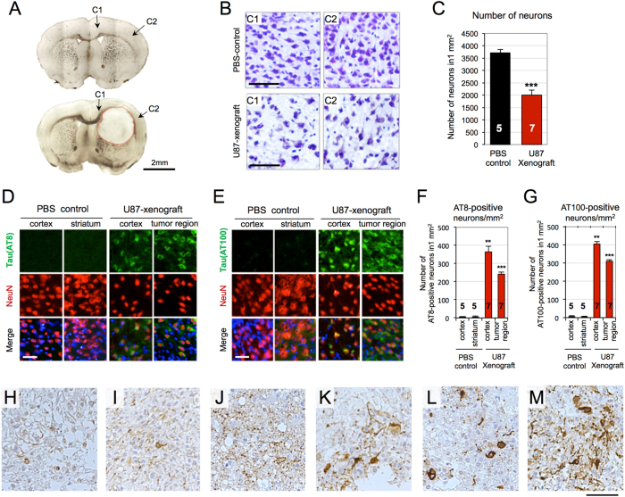
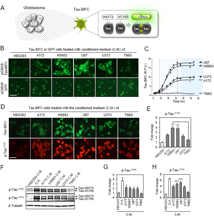
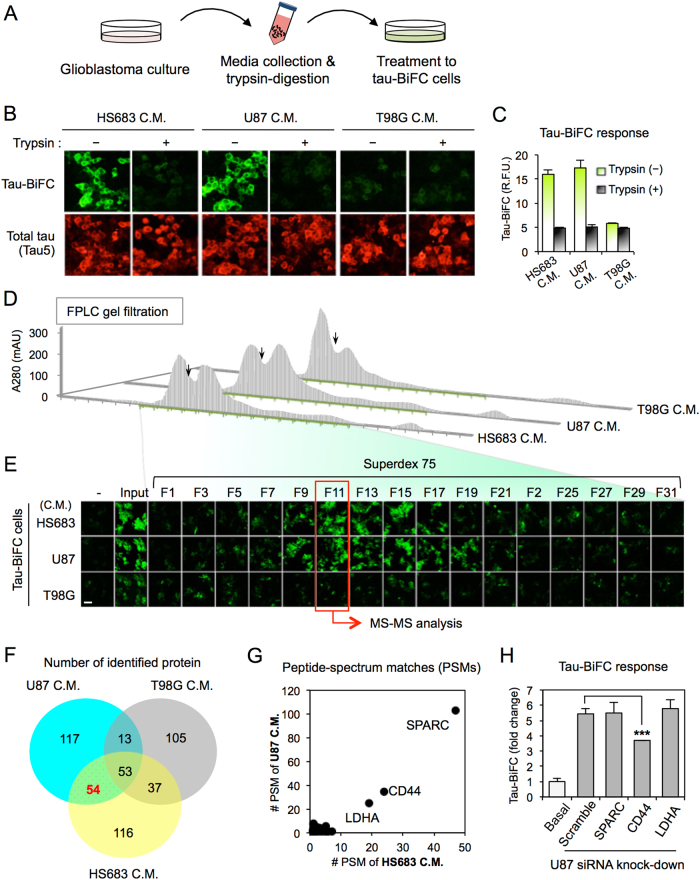
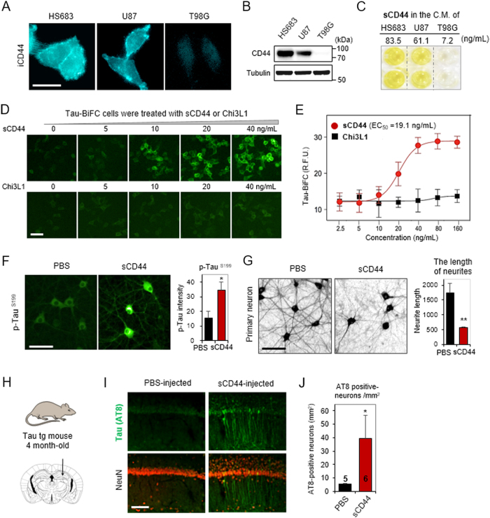
During aggressive tumor growth and migration, glioblastoma cells secrete diverse molecules and adhesion proteins to the extracellular matrix. Yet, the biochemical effects of the glioblastoma secretome in the brain remain largely unknown. Here we show that soluble CD44 secreted from glioblastoma cells induces neuronal degeneration through the activation of tau pathology in the brain. Glioblastoma-xenograft tissues showed a number of degenerating neurons bearing highly phosphorylated tau. Through a series of secretome-analyses, we identified that soluble CD44 was the responsible protein inducing tau phosphorylation and aggregation (EC = 19.1 ng/mL). The treatment of sCD44 to primary hippocampal neurons-induced tau hyperphosphorylation, leading to neuronal degeneration. Also, the injection of sCD44 into the brains of tau transgenic mice induced tau hyper-phosphorylation in hippocampal neurons. Altogether, our data suggest a neurodegenerative role of sCD44 in promoting tau pathology and serving as a molecular link between glioblastoma and neurodegeneration.

摘要

在侵袭性肿瘤生长和迁移过程中,胶质母细胞瘤细胞向细胞外基质分泌多种分子和黏附蛋白。然而,胶质母细胞瘤分泌组在大脑中的生化作用在很大程度上仍是未知的。在这里,我们表明,从胶质母细胞瘤细胞分泌的可溶性 CD44 通过激活大脑中的 tau 病理学诱导神经元变性。胶质母细胞瘤异种移植物组织显示出许多具有高度磷酸化 tau 的变性神经元。通过一系列分泌组分析,我们鉴定出可溶性 CD44 是诱导 tau 磷酸化和聚集的负责蛋白(EC = 19.1ng/mL)。sCD44 处理原代海马神经元诱导 tau 过度磷酸化,导致神经元变性。此外,将 sCD44 注射到 tau 转基因小鼠的大脑中诱导海马神经元中的 tau 过度磷酸化。总之,我们的数据表明 sCD44 在促进 tau 病理学方面具有神经退行性作用,并作为胶质母细胞瘤和神经退行性变之间的分子联系。

https://cdn.ncbi.nlm.nih.gov/pmc/blobs/9ac4/5938049/d1a4d8410a1a/12276_2017_8_Fig5_HTML.jpg
https://cdn.ncbi.nlm.nih.gov/pmc/blobs/9ac4/5938049/db0cc7a83354/12276_2017_8_Fig1_HTML.jpg
https://cdn.ncbi.nlm.nih.gov/pmc/blobs/9ac4/5938049/f604c6217c7e/12276_2017_8_Fig2_HTML.jpg
https://cdn.ncbi.nlm.nih.gov/pmc/blobs/9ac4/5938049/a8e3a66052d9/12276_2017_8_Fig3_HTML.jpg
https://cdn.ncbi.nlm.nih.gov/pmc/blobs/9ac4/5938049/4af96a2826aa/12276_2017_8_Fig4_HTML.jpg
https://cdn.ncbi.nlm.nih.gov/pmc/blobs/9ac4/5938049/d1a4d8410a1a/12276_2017_8_Fig5_HTML.jpg

相似文献

1
Glioblastoma-secreted soluble CD44 activates tau pathology in the brain.胶质母细胞瘤分泌的可溶性 CD44 激活大脑中的 tau 病理。
Exp Mol Med. 2018 Apr 6;50(4):1-11. doi: 10.1038/s12276-017-0008-7.
2
Cdk5 increases MARK4 activity and augments pathological tau accumulation and toxicity through tau phosphorylation at Ser262.Cdk5 通过磷酸化 tau 蛋白 Ser262 增加 MARK4 的活性,并增强病理性 tau 蛋白的积累和毒性。
Hum Mol Genet. 2019 Sep 15;28(18):3062-3071. doi: 10.1093/hmg/ddz120.
3
GFP-Mutant Human Tau Transgenic Mice Develop Tauopathy Following CNS Injections of Alzheimer's Brain-Derived Pathological Tau or Synthetic Mutant Human Tau Fibrils.在向中枢神经系统注射阿尔茨海默病脑源性病理性tau蛋白或合成突变型人tau蛋白原纤维后,绿色荧光蛋白标记的突变型人tau转基因小鼠出现tau蛋白病。
J Neurosci. 2017 Nov 22;37(47):11485-11494. doi: 10.1523/JNEUROSCI.2393-17.2017. Epub 2017 Oct 6.
4
Alpha1-antichymotrypsin, an inflammatory protein overexpressed in Alzheimer's disease brain, induces tau phosphorylation in neurons.α1-抗糜蛋白酶是一种在阿尔茨海默病大脑中过度表达的炎症蛋白,可诱导神经元中的tau蛋白磷酸化。
Brain. 2006 Nov;129(Pt 11):3020-34. doi: 10.1093/brain/awl255. Epub 2006 Sep 20.
5
Effect of tau-pathology on charged multivesicular body protein 2b (CHMP2B).tau 病理学对带电荷的多泡体蛋白 2b(CHMP2B)的影响。
Brain Res. 2019 Mar 1;1706:224-236. doi: 10.1016/j.brainres.2018.11.008. Epub 2018 Nov 8.
6
Increase in tau tyrosine phosphorylation correlates with the formation of tau aggregates.tau蛋白酪氨酸磷酸化的增加与tau蛋白聚集体的形成相关。
Brain Res Mol Brain Res. 2005 Aug 18;138(2):135-44. doi: 10.1016/j.molbrainres.2005.04.015.
7
Neurogenesis and cell cycle-reactivated neuronal death during pathogenic tau aggregation.致病性tau蛋白聚集过程中的神经发生和细胞周期重新激活的神经元死亡
Genes Brain Behav. 2008 Feb;7 Suppl 1(1):92-100. doi: 10.1111/j.1601-183X.2007.00377.x.
8
Differential induction and spread of tau pathology in young PS19 tau transgenic mice following intracerebral injections of pathological tau from Alzheimer's disease or corticobasal degeneration brains.在年轻的PS19 tau转基因小鼠脑内注射来自阿尔茨海默病或皮质基底节变性大脑的病理性tau后,tau病理变化的差异诱导和传播。
Acta Neuropathol. 2015 Feb;129(2):221-37. doi: 10.1007/s00401-014-1373-0. Epub 2014 Dec 24.
9
Tau hyperphosphorylation and deregulation of calcineurin in mouse models of Huntington's disease.亨廷顿氏病小鼠模型中tau蛋白的过度磷酸化及钙调神经磷酸酶的失调
Hum Mol Genet. 2015 Jan 1;24(1):86-99. doi: 10.1093/hmg/ddu456. Epub 2014 Sep 8.
10
Analysis of ribosomal protein S6 baseline phosphorylation and effect of tau pathology in the murine brain and human hippocampus.核糖体蛋白S6基线磷酸化分析以及tau病理改变对小鼠脑和人类海马体的影响。
Brain Res. 2017 Mar 15;1659:121-135. doi: 10.1016/j.brainres.2017.01.016. Epub 2017 Jan 22.

引用本文的文献

1
Frequent Alzheimer's disease neuropathological change in patients with glioblastoma.胶质母细胞瘤患者中频繁出现阿尔茨海默病神经病理改变。
Neurooncol Adv. 2024 Jul 9;6(1):vdae118. doi: 10.1093/noajnl/vdae118. eCollection 2024 Jan-Dec.
2
Beneficial Effects of Echinacoside on Cognitive Impairment and Diabetes in Type 2 Diabetic db/db Mice.松果菊苷对 2 型糖尿病 db/db 小鼠认知功能障碍和糖尿病的有益作用。
Exp Clin Endocrinol Diabetes. 2024 Aug;132(8):420-430. doi: 10.1055/a-2298-4593. Epub 2024 Apr 3.
3
Coexisting Biopsy-Diagnosed Dementia and Glioblastoma.
活检诊断的痴呆症与胶质母细胞瘤共存。
Brain Sci. 2024 Jan 30;14(2):143. doi: 10.3390/brainsci14020143.
4
Glioblastoma and its treatment are associated with extensive accelerated brain aging.胶质母细胞瘤及其治疗与广泛的加速脑老化有关。
Aging Cell. 2024 Mar;23(3):e14066. doi: 10.1111/acel.14066. Epub 2024 Jan 17.
5
Compilation of reported protein changes in the brain in Alzheimer's disease.阿尔茨海默病脑内报道的蛋白质变化汇编。
Nat Commun. 2023 Jul 25;14(1):4466. doi: 10.1038/s41467-023-40208-x.
6
Ultra High-plex Spatial Proteogenomic Investigation of Giant Cell Glioblastoma Multiforme Immune Infiltrates Reveals Distinct Protein and RNA Expression Profiles.超高多重空间蛋白质基因组学研究揭示了巨细胞胶质母细胞瘤多形性免疫浸润的独特蛋白和 RNA 表达谱。
Cancer Res Commun. 2023 May 3;3(5):763-779. doi: 10.1158/2767-9764.CRC-22-0396. eCollection 2023 May.
7
CD44 expression is correlated with osteosarcoma cell progression and immune infiltration and affects the Wnt/β-catenin signaling pathway.CD44表达与骨肉瘤细胞进展和免疫浸润相关,并影响Wnt/β-连环蛋白信号通路。
J Bone Oncol. 2023 May 25;41:100487. doi: 10.1016/j.jbo.2023.100487. eCollection 2023 Aug.
8
Endothelial glycocalyx in retina, hyperglycemia, and diabetic retinopathy.视网膜内皮糖萼、高血糖与糖尿病性视网膜病变。
Am J Physiol Cell Physiol. 2023 May 1;324(5):C1061-C1077. doi: 10.1152/ajpcell.00188.2022. Epub 2023 Mar 20.
9
Methods to detect AUTOphagy-Targeting Chimera (AUTOTAC)-mediated Targeted Protein Degradation in Tauopathies.检测自噬靶向嵌合体(AUTOTAC)介导的tau蛋白病中靶向蛋白降解的方法。
Bio Protoc. 2023 Jan 20;13(2):e4594. doi: 10.21769/BioProtoc.4594.
10
Mitochondria inter-organelle relationships in cancer protein aggregation.癌症中蛋白质聚集过程中线粒体与其他细胞器的关系
Front Cell Dev Biol. 2022 Dec 19;10:1062993. doi: 10.3389/fcell.2022.1062993. eCollection 2022.