• 文献检索
  • 文档翻译
  • 深度研究
  • 学术资讯
  • Suppr Zotero 插件Zotero 插件
  • 邀请有礼
  • 套餐&价格
  • 历史记录
应用&插件
Suppr Zotero 插件Zotero 插件浏览器插件Mac 客户端Windows 客户端微信小程序
定价
高级版会员购买积分包购买API积分包
服务
文献检索文档翻译深度研究API 文档MCP 服务
关于我们
关于 Suppr公司介绍联系我们用户协议隐私条款
关注我们

Suppr 超能文献

核心技术专利:CN118964589B侵权必究
粤ICP备2023148730 号-1Suppr @ 2026

文献检索

告别复杂PubMed语法,用中文像聊天一样搜索,搜遍4000万医学文献。AI智能推荐,让科研检索更轻松。

立即免费搜索

文件翻译

保留排版,准确专业,支持PDF/Word/PPT等文件格式,支持 12+语言互译。

免费翻译文档

深度研究

AI帮你快速写综述,25分钟生成高质量综述,智能提取关键信息,辅助科研写作。

立即免费体验

角膜穿孔合并栗子刺致晶状体损伤病例眼前节的光学相干断层扫描检查

Optical Coherence Tomography Examination of the Anterior Segment in a Case of Corneal Perforation and Lens Trauma by Chestnut Burr.

作者信息

Ono Takashi, Mori Yosai, Nejima Ryohei, Iwasaki Takuya, Amano Shiro, Miyata Kazunori

机构信息

Miyata Eye Hospital, Miyazaki, Japan.

Inouye Eye Hospital, Tokyo, Japan.

出版信息

Case Rep Ophthalmol. 2018 Feb 14;9(1):154-159. doi: 10.1159/000487076. eCollection 2018 Jan-Apr.

DOI:10.1159/000487076
PMID:29643799
原文链接:https://pmc.ncbi.nlm.nih.gov/articles/PMC5892341/
Abstract

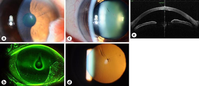
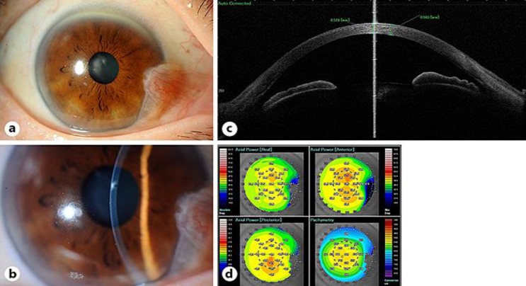
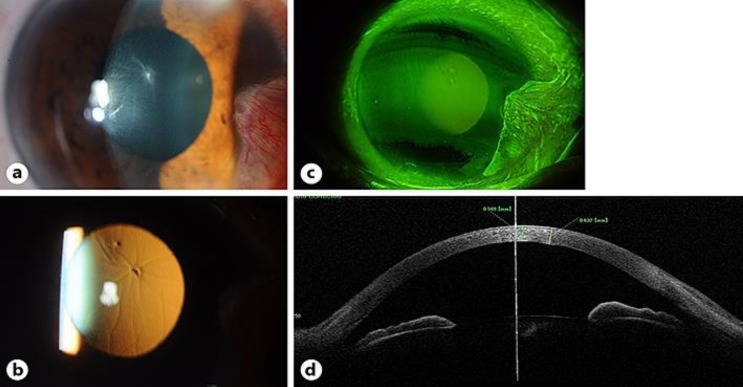
Chestnut burrs, the thorny encapsulation of chestnut fruit, can sometimes cause corneal injuries and ulceration, with poor prognoses. We report a case of corneal perforation and damaged anterior lens capsule due to a chestnut burr, using anterior segment optical coherence tomography (AS-OCT). A 67-year-old woman with a chestnut burr injury in her right eye was referred to our hospital. Her right best-corrected visual acuity (BCVA) was 0.8. Slit-lamp examination and AS-OCT showed perforation involving the endothelial layer at the center of the cornea. The iris and anterior lens capsule were damaged. Cell infiltration was observed around the wound. Bacterial examination showed gram-positive cocci but no fungi. The patient was diagnosed with a corneal perforation and bacterial keratitis. Levofloxacin 1.5% and cefmenoxime treatments were initiated and a soft contact lens was placed to seal the wound. On day 3, there was no improvement in the corneal cell infiltration, but AS-OCT suggested that the inner wound had closed. A culture test revealed the presence of , which was sensitive to both levofloxacin and cefmenoxime. Therefore, we continued the same antibiotic treatment. On day 26, the opacification and cell infiltration at the center of the cornea had improved. AS-OCT showed healing of the corneal wound with reduction in the central corneal thickness. Her BCVA improved to 1.0. AS-OCT was a valuable tool to noninvasively observe wound shape and detect the presence of any intracorneal foreign bodies.

摘要

板栗刺,即板栗果实的带刺外壳,有时会导致角膜损伤和溃疡,预后较差。我们报告一例因板栗刺导致角膜穿孔和晶状体前囊受损的病例,使用了眼前节光学相干断层扫描(AS-OCT)。一名67岁右眼被板栗刺刺伤的女性被转诊至我院。她的右眼最佳矫正视力(BCVA)为0.8。裂隙灯检查和AS-OCT显示角膜中央的穿孔累及内皮层。虹膜和晶状体前囊受损。伤口周围观察到细胞浸润。细菌检查显示革兰氏阳性球菌但无真菌。该患者被诊断为角膜穿孔和细菌性角膜炎。开始使用1.5%左氧氟沙星和头孢甲肟治疗,并放置软性接触镜以封闭伤口。第3天,角膜细胞浸润无改善,但AS-OCT提示内部伤口已闭合。培养试验显示存在[此处原文缺失具体细菌名称],其对左氧氟沙星和头孢甲肟均敏感。因此,我们继续相同的抗生素治疗。第26天,角膜中央的混浊和细胞浸润有所改善。AS-OCT显示角膜伤口愈合,中央角膜厚度减小。她的BCVA提高到1.0。AS-OCT是一种无创观察伤口形状和检测角膜内任何异物存在的有价值工具。

https://cdn.ncbi.nlm.nih.gov/pmc/blobs/f8be/5892341/de642098d3c3/cop-0009-0154-g03.jpg
https://cdn.ncbi.nlm.nih.gov/pmc/blobs/f8be/5892341/272b2f2c9517/cop-0009-0154-g01.jpg
https://cdn.ncbi.nlm.nih.gov/pmc/blobs/f8be/5892341/9af1754b6aac/cop-0009-0154-g02.jpg
https://cdn.ncbi.nlm.nih.gov/pmc/blobs/f8be/5892341/de642098d3c3/cop-0009-0154-g03.jpg
https://cdn.ncbi.nlm.nih.gov/pmc/blobs/f8be/5892341/272b2f2c9517/cop-0009-0154-g01.jpg
https://cdn.ncbi.nlm.nih.gov/pmc/blobs/f8be/5892341/9af1754b6aac/cop-0009-0154-g02.jpg
https://cdn.ncbi.nlm.nih.gov/pmc/blobs/f8be/5892341/de642098d3c3/cop-0009-0154-g03.jpg

相似文献

1
Optical Coherence Tomography Examination of the Anterior Segment in a Case of Corneal Perforation and Lens Trauma by Chestnut Burr.角膜穿孔合并栗子刺致晶状体损伤病例眼前节的光学相干断层扫描检查
Case Rep Ophthalmol. 2018 Feb 14;9(1):154-159. doi: 10.1159/000487076. eCollection 2018 Jan-Apr.
2
Case Report: Evaluating Intraocular Foreign Bodies After Corneal Perforation Using Swept Source Anterior Segment Optical Coherence Tomography.病例报告:使用扫频源眼前节光学相干断层扫描评估角膜穿孔后的眼内异物
Optom Vis Sci. 2020 Feb;97(2):101-103. doi: 10.1097/OPX.0000000000001475.
3
Controversy of indomethacin eye drops in the treatment of rheumatoid arthritis-induced corneal ulceration: a case report.吲哚美辛滴眼液治疗类风湿关节炎相关性角膜溃疡的争议:一例报告。
J Med Case Rep. 2021 Mar 4;15(1):116. doi: 10.1186/s13256-020-02600-9.
4
Femtosecond laser-assisted removal of an intracorneal chestnut, a case report.飞秒激光辅助摘除角膜内异物,病例报告
BMC Ophthalmol. 2018 Aug 28;18(1):210. doi: 10.1186/s12886-018-0875-2.
5
Wound healing process after corneal stromal thinning observed with anterior segment optical coherence tomography.眼前节光学相干断层扫描观察角膜基质变薄后的伤口愈合过程。
Cornea. 2014 Oct;33(10):1056-60. doi: 10.1097/ICO.0000000000000223.
6
Retrospective Evaluation of Corneal Foreign Bodies with Anterior Segment Optical Coherence Tomography.眼前节光学相干断层扫描对角膜异物的回顾性评估。
Turk J Ophthalmol. 2021 Oct 26;51(5):265-268. doi: 10.4274/tjo.galenos.2020.42223.
7
Chronically retained feathery chestnut-burr spine-related corneal injury: clinical features and outcome.慢性滞留的羽扇豆状栗刺相关角膜损伤:临床特征和结局。
Int Ophthalmol. 2020 Aug;40(8):1993-1997. doi: 10.1007/s10792-020-01374-9. Epub 2020 Apr 23.
8
[New diagnostic methods for imaging the anterior segment of the eye to enable treatment modalities selection].[用于眼部前段成像以辅助治疗方式选择的新诊断方法]
Nippon Ganka Gakkai Zasshi. 2011 Mar;115(3):297-322; discussion 323.
9
The role of anterior segment optical coherence tomography in the management of an intra-corneal foreign body.眼前节光学相干断层扫描在角膜异物处理中的作用
Springerplus. 2016 Sep 13;5(1):1559. doi: 10.1186/s40064-016-3242-x. eCollection 2016.
10
[Clinical observation of ocular injury caused by chestnut burr].[板栗毛刺致眼外伤的临床观察]
Zhonghua Yan Ke Za Zhi. 2020 May 11;56(5):370-375. doi: 10.3760/cma.j.cn112142-20190604-00295.

引用本文的文献

1
Management of intrascleral chestnut burr spines under ultrasound biomicroscopy guidance.超声生物显微镜引导下巩膜内栗刺状异物的处理
Indian J Ophthalmol. 2022 Sep;70(9):3311-3315. doi: 10.4103/ijo.IJO_356_22.
2
Clinical outcome after air-assisted manual deep anterior lamellar keratoplasty for fungal keratitis poorly responsive to medical treatment.空气辅助手动深板层角膜移植术治疗药物治疗效果不佳的真菌性角膜炎后的临床结果。
Clin Ophthalmol. 2019 Sep 26;13:1913-1919. doi: 10.2147/OPTH.S211099. eCollection 2019.
3
Corneal injury: Clinical and molecular aspects.

本文引用的文献

1
The role of anterior segment optical coherence tomography in the management of an intra-corneal foreign body.眼前节光学相干断层扫描在角膜异物处理中的作用
Springerplus. 2016 Sep 13;5(1):1559. doi: 10.1186/s40064-016-3242-x. eCollection 2016.
2
Ocular Injury With Chestnut Burr: Our Experience.板栗毛刺致眼外伤:我们的经验
Cornea. 2016 Oct;35(10):1315-9. doi: 10.1097/ICO.0000000000000952.
3
A 7-year retrospective study for clinical features and visual outcome of chestnut burr-related ocular injuries.一项关于板栗毛刺相关眼外伤临床特征及视力预后的7年回顾性研究。
角膜损伤:临床与分子方面。
Exp Eye Res. 2019 Sep;186:107709. doi: 10.1016/j.exer.2019.107709. Epub 2019 Jun 22.
Graefes Arch Clin Exp Ophthalmol. 2013 Apr;251(4):1247-9. doi: 10.1007/s00417-012-2104-7. Epub 2012 Jul 22.
4
[Ocular mycosis].[眼部真菌病]
Nihon Ishinkin Gakkai Zasshi. 2008;49(3):175-9. doi: 10.3314/jjmm.49.175.
5
A system for classifying mechanical injuries of the eye (globe). The Ocular Trauma Classification Group.一种用于对眼球机械性损伤进行分类的系统。眼外伤分类小组。
Am J Ophthalmol. 1997 Jun;123(6):820-31. doi: 10.1016/s0002-9394(14)71132-8.
6
Paddy grain injury of the eye. A study of 277 cases.稻谷致眼损伤。277例病例研究。
Br J Ophthalmol. 1968 Feb;52(2):191-7. doi: 10.1136/bjo.52.2.191.