Department of Pediatrics, Maastricht University Medical Center, 6202, AZ, Maastricht, The Netherlands.
School for Mental Health and Neuroscience (MHeNs), Maastricht University Medical Center, 6229, ER, Maastricht, The Netherlands.
J Neuroinflammation. 2018 Apr 19;15(1):113. doi: 10.1186/s12974-018-1149-x.
Antenatal infection (i.e., chorioamnionitis) is an important risk factor for adverse neurodevelopmental outcomes after preterm birth. Destructive and developmental disturbances of the white matter are hallmarks of preterm brain injury. Understanding the temporal effects of antenatal infection in relation to the onset of neurological injury is crucial for the development of neurotherapeutics for preterm infants. However, these dynamics remain unstudied.
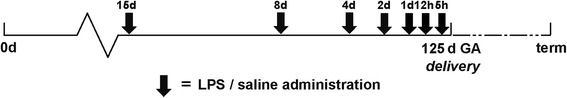
Time-mated ewes were intra-amniotically injected with lipopolysaccharide at 5, 12, or 24 h or 2, 4, 8, or 15 days before preterm delivery at 125 days gestational age (term ~ 150 days). Post mortem analyses for peripheral immune activation, neuroinflammation, and white matter/neuronal injury were performed. Moreover, considering the neuroprotective potential of erythropoietin (EPO) for perinatal brain injury, we evaluated (phosphorylated) EPO receptor (pEPOR) expression in the fetal brain following LPS exposure.
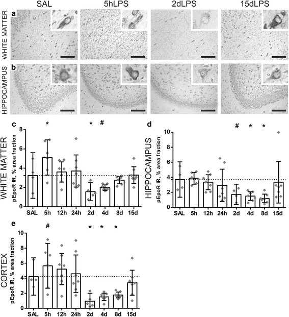
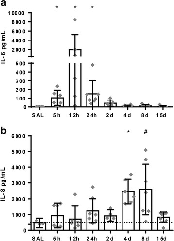
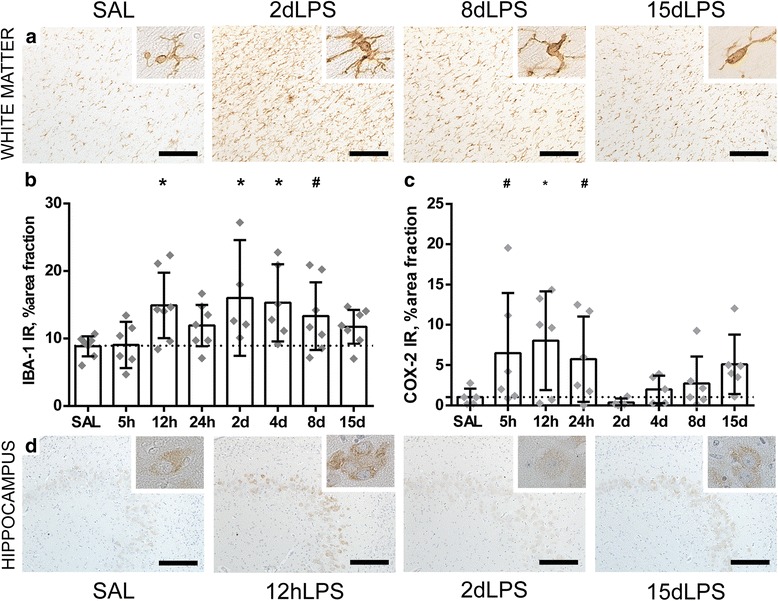
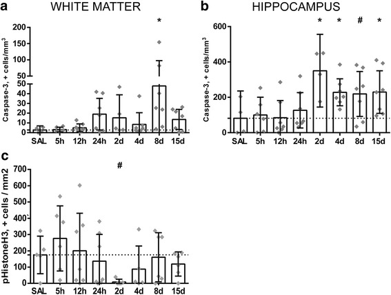
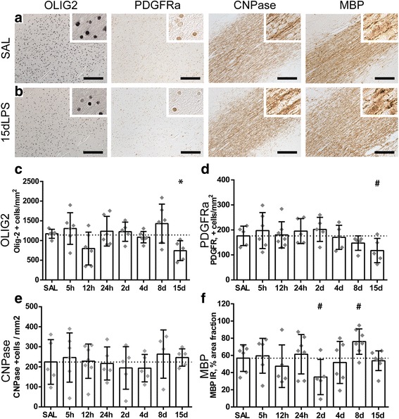
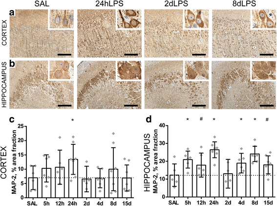
Intra-amniotic exposure to this single bolus of LPS resulted in a biphasic systemic IL-6 and IL-8 response. In the developing brain, intra-amniotic LPS exposure induces a persistent microgliosis (IBA-1 immunoreactivity) but a shorter-lived increase in the pro-inflammatory marker COX-2. Cell death (caspase-3 immunoreactivity) was only observed when LPS exposure was greater than 8 days in the white matter, and there was a reduction in the number of (pre) oligodendrocytes (Olig2- and PDGFRα-positive cells) within the white matter at 15 days post LPS exposure only. pEPOR expression displayed a striking biphasic regulation following LPS exposure which may help explain contradicting results among clinical trials that tested EPO for the prevention of preterm brain injury.
We provide increased understanding of the spatiotemporal pathophysiological changes in the preterm brain following intra-amniotic inflammation which may aid development of new interventions or implement interventions more effectively to prevent perinatal brain damage.
产前感染(即绒毛膜羊膜炎)是早产儿发生不良神经发育结局的重要危险因素。早产儿脑损伤的特征是白质的破坏性和发育性紊乱。了解产前感染与神经损伤发作的时间关系对于开发早产儿神经治疗方法至关重要。然而,这些动态尚未得到研究。
时间匹配的母羊在 125 天妊娠龄(足月~150 天)前,于早产前 5、12 或 24 小时或 2、4、8 或 15 天经羊膜内注射脂多糖。进行外周免疫激活、神经炎症和白质/神经元损伤的死后分析。此外,考虑到促红细胞生成素(EPO)对围产期脑损伤的神经保护潜力,我们评估了 LPS 暴露后胎儿脑中(磷酸化)EPO 受体(pEPOR)的表达。
单次羊膜内 LPS 暴露导致系统中 IL-6 和 IL-8 的双相反应。在发育中的大脑中,羊膜内 LPS 暴露会引起持续的小胶质细胞增生(IBA-1 免疫反应),但促炎标志物 COX-2 的短暂增加。只有在 LPS 暴露超过 8 天时,才会在白质中观察到细胞死亡(caspase-3 免疫反应),并且只有在 LPS 暴露后 15 天,白质内(前)少突胶质细胞(Olig2- 和 PDGFRα 阳性细胞)的数量才会减少。pEPOR 表达在 LPS 暴露后表现出明显的双相调节,这可能有助于解释临床试验中测试 EPO 预防早产儿脑损伤的结果相互矛盾的原因。
我们提供了对羊膜内炎症后早产儿脑的时空病理生理变化的更深入了解,这可能有助于开发新的干预措施或更有效地实施干预措施,以预防围产期脑损伤。