Department of Ophthalmology, Haeundae Paik Hospital, Inje University College of Medicine, Busan, South Korea.
Hamilton Glaucoma Center, Shiley Eye Institute, the Department of Ophthalmology, University of California San Diego, La Jolla, California, United States.
Invest Ophthalmol Vis Sci. 2018 Apr 1;59(5):1995-2004. doi: 10.1167/iovs.17-23046.
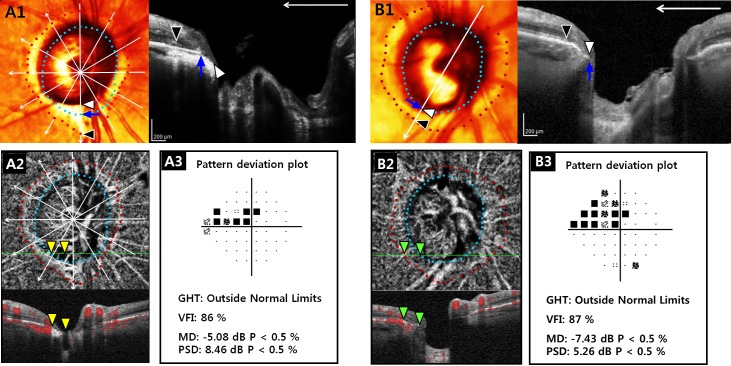
To investigate the association between the microstructure of β-zone parapapillary atrophy (βPPA) and parapapillary deep-layer microvasculature dropout assessed by optical coherence tomography angiography (OCT-A).
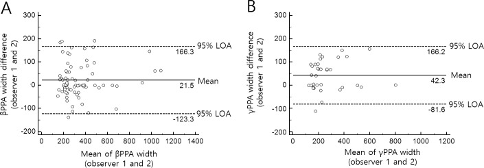
Thirty-seven eyes with βPPA devoid of the Bruch's membrane (BM) (γPPA) ranging between completely absent and discontinuous BM were matched by severity of the visual field (VF) damage with 37 eyes with fully intact BM (βPPA+BM) based on the spectral-domain (SD) OCT imaging. Parapapillary deep-layer microvasculature dropout was defined as a dropout of the microvasculature within choroid or scleral flange in the βPPA on the OCT-A. The widths of βPPA, γPPA, and βPPA+BM were measured on six radial SD-OCT images. Prevalence of the dropout was compared between eyes with and without γPPA. Logistic regression was performed for evaluating association of the dropout with the width of βPPA, γPPA, and βPPA+BM, and the γPPA presence.
Eyes with γPPA had significantly higher prevalence of the dropout than did those without γPPA (75.7% versus 40.8%; P = 0.004). In logistic regression, presence and longer width of the γPPA, worse VF mean deviation, and presence of focal lamina cribrosa defects were significantly associated with the dropout (P < 0.05), whereas width of the βPPA and βPPA+BM, axial length, and choroidal thickness were not (P > 0.10).
Parapapillary deep-layer microvasculature dropout was associated with the presence and larger width of γPPA, but not with the βPPA+BM width. Presence and width of the exposed scleral flange, rather than the retinal pigmented epithelium atrophy, may be associated with deep-layer microvasculature dropout.
探讨光学相干断层扫描血管造影(OCTA)评估的β-区视盘旁萎缩(βPPA)的微观结构与视盘旁深层微血管丢失之间的关系。
根据光谱域(SD)OCT 成像,将 37 只完全无(γPPA)或不连续 BM 的βPPA (γPPA)与 37 只完全有 BM 的βPPA(βPPA+BM)进行匹配,匹配标准为视野(VF)损害的严重程度。视盘旁深层微血管丢失定义为 OCT-A 上βPPA 中脉络膜或巩膜边缘的微血管丢失。在 6 张径向 SD-OCT 图像上测量βPPA、γPPA 和βPPA+BM 的宽度。比较有γPPA 和无γPPA 眼之间的丢失率。采用 Logistic 回归评估丢失与βPPA、γPPA 和βPPA+BM 宽度以及γPPA 存在的相关性。
有γPPA 的眼比无γPPA 的眼丢失率更高(75.7%比 40.8%;P=0.004)。Logistic 回归显示,γPPA 的存在和更长的宽度、更差的 VF 平均偏差以及焦点筛板缺陷的存在与丢失显著相关(P<0.05),而βPPA 和βPPA+BM 的宽度、眼轴长度和脉络膜厚度与丢失无关(P>0.10)。
视盘旁深层微血管丢失与γPPA 的存在和较大宽度相关,与βPPA+BM 宽度无关。暴露的巩膜边缘的存在和宽度,而不是视网膜色素上皮萎缩,可能与深层微血管丢失相关。