Rehman Lal, Farooq Ghulam, Bukhari Irum
Department of Neurosurgery, Jinnah Postgraduate Medical Center, Karachi, Pakistan.
Asian J Neurosurg. 2018 Apr-Jun;13(2):233-237. doi: 10.4103/1793-5482.228549.
The aim of this study is to find the outcome of repair and resection of the occipital encephalocele.
Case series.
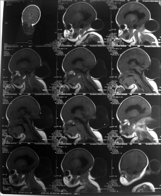
The clinical data of fifty consecutive occipital encephalocele patients were retrieved from medical records including operative notes, postoperative follow-up visits, and postsurgical complications were noted for analysis from November 2009 to November 2013 at the Department of Neurosurgery, Jinnah Postgraduate Medical Centre, Karachi, Pakistan. All patients were assessed by computed tomography scan, magnetic resonance imaging brain, and ultrasound when needed. Physician's assessment, physical examination, and his/her questions to the family at follow-up were used as a tool to determine if there was a developmental delay rather than quantitative analysis like hydrocephalus questionnaires. Patients who developed complications and delayed milestone were regarded as no improvement and those who did not develop complications and achieved appropriate milestone were regarded as improved at 18 months follow-up.
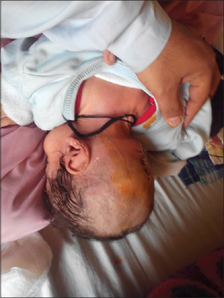
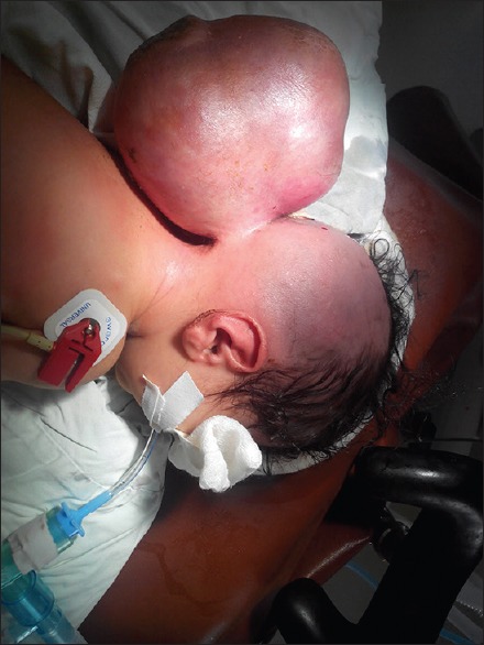
Of 50 patients, 17 were males and 33 were females. The average age at presentation was 2.4 months. 16 (32%) patients had increased head circumference and hydrocephalus, 2 (4%) had associated Dandy-Walker cyst, 3 (6%) developed developmental delays, and 8 (15%) had a seizure disorder. None of our patients had neurological deficits. The size of the sac ranged from 2 cm × 3 cm to 27 cm × 15 cm. 9 (18%) patients were admitted with the complication of sac rupture and 2 (4%) patients sac ruptured after admission. Only one patient (2%) had a cerebrospinal fluid leak postoperatively that was repaired primarily without patch graft or dura seal while 4 (8%) developed hydrocephalus after repair of the sac which was treated with placement of ventriculoperitoneal shunt. One (2%) patient did not recover from anesthesia and expired.
Encephalocele is commonly seen in the practice of neurosurgery in the world as well as in Pakistan. Modern neuroimaging, neurosurgical techniques, and neonatal neurological intensive care have greatly improved morbidity and mortality in the care of encephalocele.
本研究的目的是找出枕部脑膨出修补和切除的结果。
病例系列。
从2009年11月至2013年11月在巴基斯坦卡拉奇真纳研究生医学中心神经外科的病历中检索了50例连续枕部脑膨出患者的临床资料,包括手术记录、术后随访,并记录术后并发症以进行分析。所有患者在需要时均接受计算机断层扫描、脑部磁共振成像和超声检查。医生的评估、体格检查以及随访时向家属提出的问题被用作确定是否存在发育迟缓的工具,而非像脑积水问卷那样进行定量分析。在18个月的随访中,出现并发症和发育里程碑延迟的患者被视为无改善,而未出现并发症且达到适当发育里程碑的患者被视为改善。
50例患者中,男性17例,女性33例。就诊时的平均年龄为2.4个月。16例(32%)患者头围增大并伴有脑积水,2例(4%)伴有丹迪-沃克囊肿,3例(6%)出现发育迟缓,8例(15%)患有癫痫症。我们的患者均无神经功能缺损。囊的大小范围为2厘米×3厘米至27厘米×15厘米。9例(18%)患者因囊破裂并发症入院,2例(4%)患者入院后囊破裂。只有1例患者(2%)术后出现脑脊液漏,在未使用补片移植或硬脑膜封闭的情况下进行了一期修复,而4例(8%)患者在囊修复后出现脑积水,通过放置脑室腹腔分流管进行治疗。1例(2%)患者未从麻醉中苏醒并死亡。
脑膨出在世界以及巴基斯坦的神经外科实践中都很常见。现代神经影像学、神经外科技术和新生儿神经重症监护极大地改善了脑膨出治疗中的发病率和死亡率。