Institute for Cancer Genetics and Informatics, Oslo University Hospital, Oslo, Norway.
Center for Cancer Biomedicine, University of Oslo, Oslo, Norway.
J Natl Cancer Inst. 2018 Dec 1;110(12):1400-1408. doi: 10.1093/jnci/djy063.
Nuclear texture analysis measuring differences in chromatin structure has provided prognostic biomarkers in several cancers. There is a need for improved cell-by-cell chromatin analysis to detect nuclei with highly disorganized chromatin. The purpose of this study was to develop a method for detecting nuclei with high chromatin entropy and to evaluate the association between the presence of such deviating nuclei and prognosis.
A new texture-based biomarker that characterizes each cancer based on the proportion of high-chromatin entropy nuclei (<25% vs ≥25%) was developed on a discovery set of 175 uterine sarcomas. The prognostic impact of this biomarker was evaluated on a validation set of 179 uterine sarcomas, as well as on independent validation sets of 246 early-stage ovarian carcinomas and 791 endometrial carcinomas. More than 1 million images of nuclei stained for DNA were included in the study. All statistical tests were two-sided.
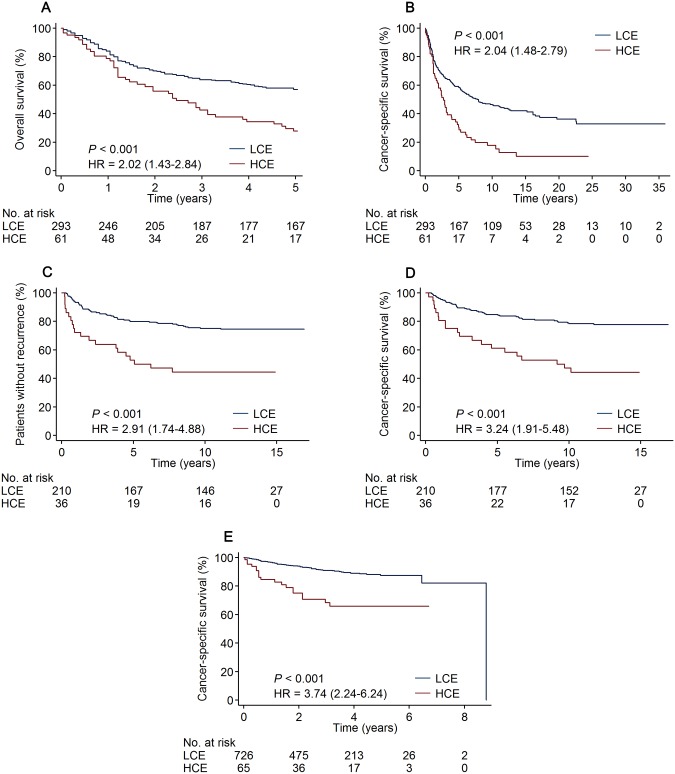
An increased proportion of high-chromatin entropy nuclei was associated with poor clinical outcome. The biomarker predicted five-year overall survival for uterine sarcoma patients with a hazard ratio (HR) of 2.02 (95% confidence interval [CI] = 1.43 to 2.84), time to recurrence for ovarian cancer patients (HR = 2.91, 95% CI = 1.74 to 4.88), and cancer-specific survival for endometrial cancer patients (HR = 3.74, 95% CI = 2.24 to 6.24). Chromatin entropy was an independent prognostic marker in multivariable analyses with clinicopathological parameters (HR = 1.81, 95% CI = 1.21 to 2.70, for sarcoma; HR = 1.71, 95% CI = 1.01 to 2.90, for ovarian cancer; and HR = 2.03, 95% CI = 1.19 to 3.45, for endometrial cancer).
A novel method detected high-chromatin entropy nuclei, and an increased proportion of such nuclei was associated with poor prognosis. Chromatin entropy supplemented existing prognostic markers in multivariable analyses of three gynecological cancer cohorts.
核纹理分析测量染色质结构的差异为几种癌症提供了预后生物标志物。需要进行改进的细胞间染色质分析,以检测具有高度紊乱染色质的核。本研究的目的是开发一种检测高染色质熵核的方法,并评估存在这种偏离核与预后之间的关系。
在 175 例子宫肉瘤的发现组中开发了一种基于新的基于纹理的生物标志物,该标志物根据高染色质熵核(<25%与≥25%)的比例对每个癌症进行特征描述。在 179 例子宫肉瘤的验证组以及 246 例早期卵巢癌和 791 例子宫内膜癌的独立验证组中评估了该生物标志物的预后影响。研究中包含了超过 100 万张用于 DNA 染色的核图像。所有统计检验均为双侧检验。
高染色质熵核的比例增加与临床结局不良相关。该生物标志物预测了子宫肉瘤患者的五年总生存率,风险比(HR)为 2.02(95%置信区间[CI]为 1.43 至 2.84),卵巢癌患者的复发时间(HR=2.91,95%CI=1.74 至 4.88),以及子宫内膜癌患者的癌症特异性生存率(HR=3.74,95%CI=2.24 至 6.24)。在多变量分析中,染色质熵是与临床病理参数独立的预后标志物(HR=1.81,95%CI=1.21 至 2.70,用于肉瘤;HR=1.71,95%CI=1.01 至 2.90,用于卵巢癌;HR=2.03,95%CI=1.19 至 3.45,用于子宫内膜癌)。
一种新方法检测到高染色质熵核,并且这种核的比例增加与预后不良相关。染色质熵在三个妇科癌症队列的多变量分析中补充了现有的预后标志物。