Sun Li-Li, Li Zhong-Hao, Tang Wen-Xiong, Liu Lei, Chang Fei-Yan, Zhang Xue-Bin, Ye Wei-Jie, Lu Shuo, Liu Zun-Jing, Zhu Xian-Jin
Department of Neurology, China-Japan Friendship Hospital, 2 Yinghua Dongjie, Hepingli, Beijing, 100029, China.
Department of Radiology, China-Japan Friendship Hospital, 2 Yinghua Dongjie, Hepingli, Beijing, 100029, China.
BMC Neurol. 2018 Apr 25;18(1):51. doi: 10.1186/s12883-018-1054-z.
It is usually difficult to identify stroke pathogenesis for single lenticulostriate infarction with nonstenotic middle cerebral artery (MCA). Our aim is to differentiate the two pathogeneses, non-branch atheromatous small vessel disease and branch atheromatous disease (BAD) by high-resolution magnetic resonance imaging (HR-MRI).
Thirty-two single lenticulostriate infarction patients with nonstenotic MCA admitted to the China-Japan Friendship Hospital from December 2014 to August 2017 were enrolled for retrospective analysis. National Institutes of Health Stroke Scale (NIHSS), modified Rankin Scale (mRS), atherosclerotic risk factors, imaging features, and the characteristic of MCA vessel wall in HR-MRI were evaluated.
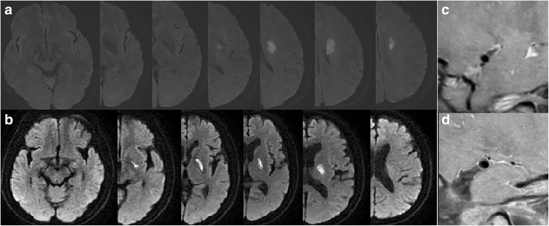
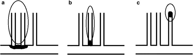
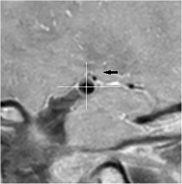
MCA plaques were detected in 15(46.9%) patients which implied BAD and 8 of 15 (53.3%) patients had plaques location in upper dorsal side of the vessel wall. Patients with HR-MRI identified plaques had a significantly larger infarction lesion length (1.95 ± 0.86 cm versus 1.38 ± 0.55 cm; P = 0.031) and larger lesion volume (2.95 ± 3.94 cm versus 0.90 ± 0.94 cm; P = 0.027) than patients without plaques. Patients with HR-MRI identified plaques had a significant higher percentage of proximal lesions than patients without plaques (P = 0.055). However, according to the location of MCA plaques, there were no significant differences in terms of imaging features, NIHSS and mRS.
We demonstrated high frequency of MCA atheromatous plaques visualized in single lenticulostriate infarction patients with nonstenotic MCA by using HR-MRI. Patients with HR-MRI identified plaque presented larger infarction lesions and more proximal lesions than patients without plaque, which were consistent with imaging features of BAD. HR-MRI is an important and effective tool for identifying stroke etiology in patients with nonstenotic MCA.
对于大脑中动脉(MCA)无狭窄的单纯豆纹动脉梗死,通常很难确定其发病机制。我们的目的是通过高分辨率磁共振成像(HR-MRI)区分两种发病机制,即非分支动脉粥样硬化性小血管病和分支动脉粥样硬化病(BAD)。
回顾性分析2014年12月至2017年8月在中国-日本友好医院收治的32例MCA无狭窄的单纯豆纹动脉梗死患者。评估美国国立卫生研究院卒中量表(NIHSS)、改良Rankin量表(mRS)、动脉粥样硬化危险因素、影像学特征以及HR-MRI中MCA血管壁的特征。
15例(46.9%)患者检测到MCA斑块,提示BAD,其中8例(53.3%)患者的斑块位于血管壁的上背侧。HR-MRI显示有斑块的患者梗死灶长度明显长于无斑块的患者(1.95±0.86cm对1.38±0.55cm;P = 0.031),病灶体积也更大(2.95±3.94cm对0.90±0.94cm;P = 0.027)。HR-MRI显示有斑块的患者近端病灶的比例显著高于无斑块的患者(P = 0.055)。然而,根据MCA斑块的位置,在影像学特征、NIHSS和mRS方面没有显著差异。
我们通过HR-MRI证明,在MCA无狭窄的单纯豆纹动脉梗死患者中,MCA动脉粥样硬化斑块的可视化频率较高。HR-MRI显示有斑块的患者比无斑块的患者梗死灶更大且近端病灶更多,这与BAD的影像学特征一致。HR-MRI是识别MCA无狭窄患者卒中病因的重要且有效的工具。