Wen Liang, Shi Xinan, He Liping, Lu Yi, Han Dan
1Radiology department , The First Affiliated Hospital of Kunming Medical University, 295 Xichang Road, Kunming city, Yunnan province 650000 People's Republic of China.
2Kunming Medical University, Kunming City, People's Republic of China.
Eur Radiol Exp. 2017;1(1):21. doi: 10.1186/s41747-017-0024-3. Epub 2017 Nov 2.
To study manganese superoxide dismutase (MnSOD) expression, manganese-enhanced magnetic resonance imaging (MEMRI) appearance and its relation to metastatic potential in colorectal cancer (CRC).
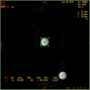
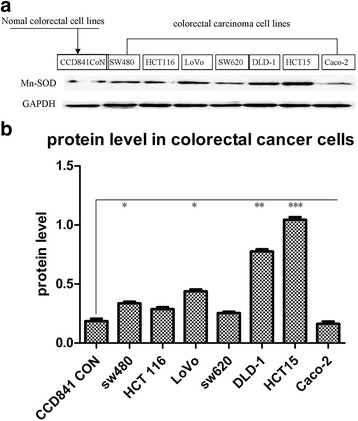
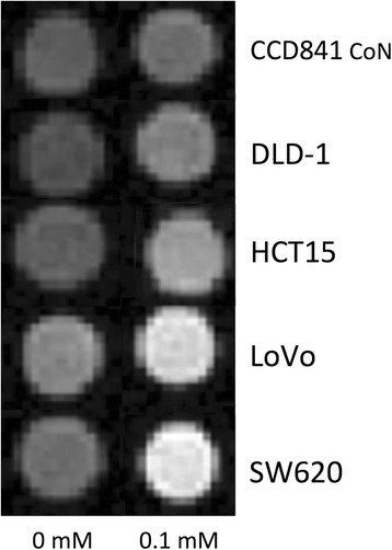
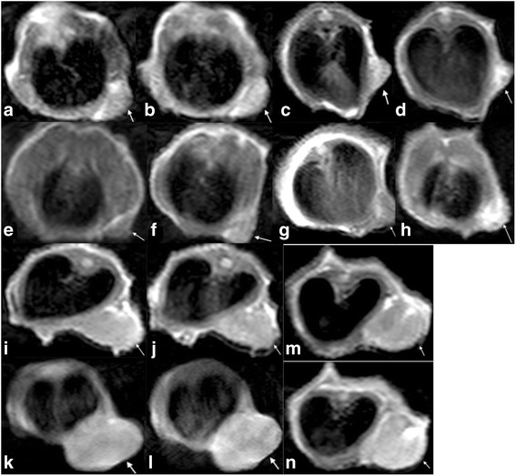
CRC cells SW620, HCT116, LoVo, SW480, DLD-1, HCT15, Caco-2 and their normal counterpart CCD841 CoN were chosen, based on differential aggressiveness, to undergo Western blot analysis for assessment of MnSOD expression, reported as proportion of readings to internal reference (glyceraldehyde-3-phosphate-dehydrogenase). Based on the results of the invasion assay, HCT15, DLD-1, LoVo and SW620 cells and corresponding xenografts underwent MEMRI. The differences of average T1-value shortening were compared.
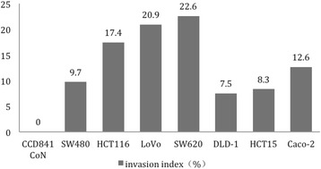
MnSOD expression in SW620, HCT116, LoVo, SW480, DLD-1, HCT15, Caco-2 and CCD841 CoN cells (0.255 ± 0.018 (mean ± standard deviation), 0.289 ± 0.028, 0.438 ± 0.028, 0.337 ± 0.025, 0.777 ± 0.031, 1.045 ± 0.038, 0.163 ± 0.035 and 0.185 ± 0.038, respectively) was not correlated with Invasion Index (22.6 ± 0.7, 17.0 ± 0.6, 20.9 ± 0.6, 9.7 ± 0.4, 7.5 ± 0.3, 8.3 ± 0.2, 12.6 ± 0.5 and 0) (r = - 0.204, = 0.627). In highly aggressive cells (SW620, LoVo), T1 shortening (289.33 ± 0.57, 268.45 ± 6.87 ms, respectively) was greater than that in lower counterparts (148.68 ± 3.99 ms in DLD-1, 128.60 ± 1.96 in HCT15) ( < 0.001). Both 5- and 10-mm group SW620 and/or LoVo tumours showed greater T1 shortening (≥600 ms) than DLD-1 and HCT15 (≤350 ms) ( < 0.001, = 0.005, = 0.010).
MEMRI has the potential to noninvasively distinguish different metastatic potential CRCs. However, the MnSOD expression is not correlated to malignant potential in CRC cells.
研究锰超氧化物歧化酶(MnSOD)表达、锰增强磁共振成像(MEMRI)表现及其与结直肠癌(CRC)转移潜能的关系。
基于侵袭性差异,选取CRC细胞SW620、HCT116、LoVo、SW480、DLD-1、HCT15、Caco-2及其正常对照CCD841 CoN,进行蛋白质免疫印迹分析以评估MnSOD表达,结果以与内参(甘油醛-3-磷酸脱氢酶)读数的比例表示。根据侵袭实验结果,对HCT15、DLD-1、LoVo和SW620细胞及其相应异种移植瘤进行MEMRI。比较平均T1值缩短的差异。
SW620、HCT116、LoVo、SW480、DLD-1、HCT15、Caco-2和CCD841 CoN细胞中的MnSOD表达(分别为0.255±0.018(均值±标准差)、0.289±0.028、0.438±0.028、0.337±0.025、0.777±0.031、1.045±0.038、0.163±0.035和0.185±0.038)与侵袭指数(22.6±0.7、17.0±0.6、20.9±0.6、9.7±0.4、7.5±0.3、8.3±0.2、12.6±0.5和0)不相关(r = -0.204,P = 0.627)。在高侵袭性细胞(SW620、LoVo)中,T1缩短(分别为289.33±0.57、268.45±6.87 ms)大于低侵袭性细胞(DLD-1中为148.68±3.99 ms,HCT15中为128.60±1.96)(P < 0.001)。5 mm和10 mm组的SW620和/或LoVo肿瘤显示出比DLD-1和HCT15更大的T1缩短(≥600 ms)(≤350 ms)(P < 0.001,P = 0.005,P = 0.010)。
MEMRI有潜力无创区分不同转移潜能的CRC。然而,MnSOD表达与CRC细胞的恶性潜能不相关。