1 Safar Center for Resuscitation Research, Department of Critical Care Medicine, University of Pittsburgh, PA, USA.
2 Pittsburgh NMR Center for Biomedical Research, Carnegie Mellon University, PA, USA.
ASN Neuro. 2018 Jan-Dec;10:1759091418770543. doi: 10.1177/1759091418770543.
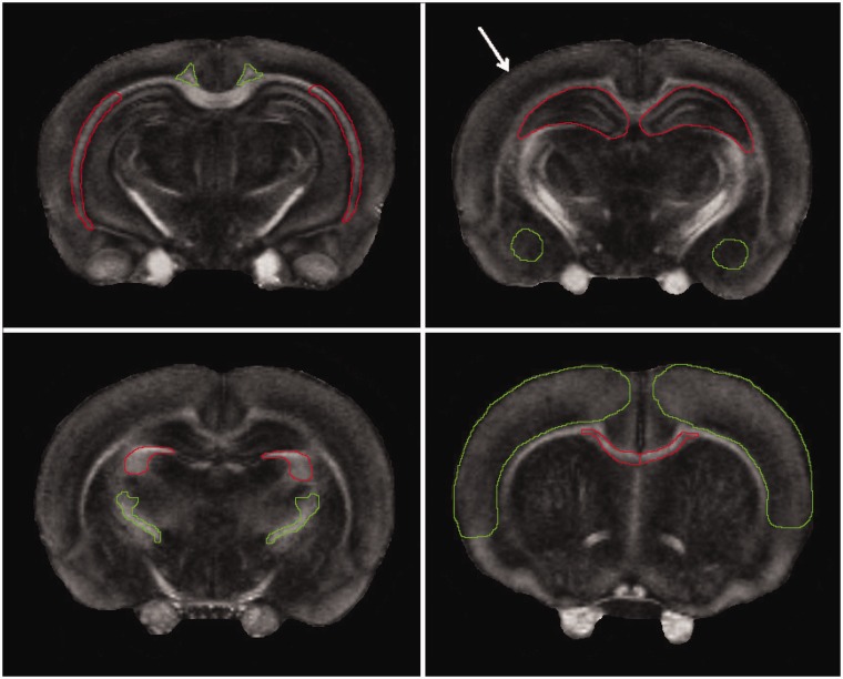
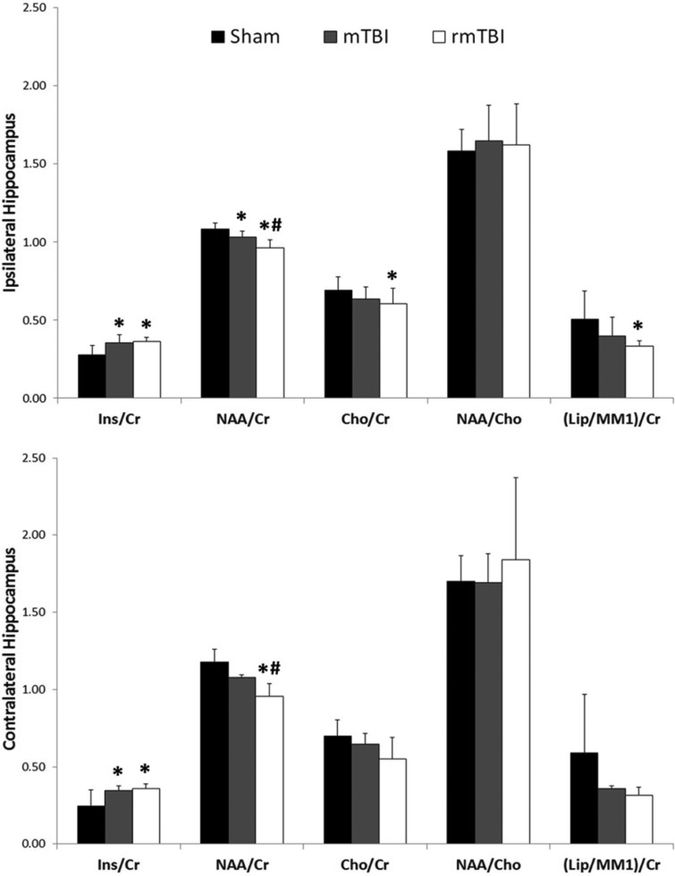
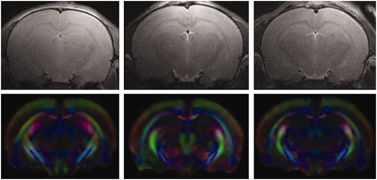
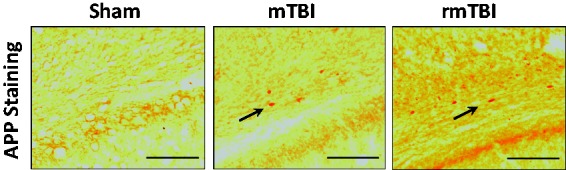
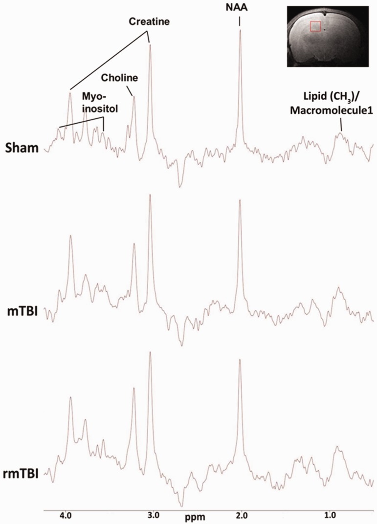
Mild traumatic brain injury (mTBI) in children is a common and serious public health problem. Traditional neuroimaging findings in children who sustain mTBI are often normal, putting them at risk for repeated mTBI (rmTBI). There is a need for more sensitive imaging techniques capable of detecting subtle neurophysiological alterations after injury. We examined neurochemical and white matter changes using diffusion tensor imaging of the whole brain and proton magnetic resonance spectroscopy of the hippocampi at 7 Tesla in 18-day-old male rats at 7 days after mTBI and rmTBI. Traumatic axonal injury was assessed by beta-amyloid precursor protein accumulation using immunohistochemistry. A significant decrease in fractional anisotropy and increase in axial and radial diffusivity were observed in several brain regions, especially in white matter regions, after a single mTBI versus sham and more prominently after rmTBI. In addition, we observed accumulation of beta-amyloid precursor protein in the external capsule after mTBI and rmTBI. mTBI and rmTBI reduced the N-acetylaspartate/creatine ratio (NAA/Cr) and increased the myoinositol/creatine ratio (Ins/Cr) versus sham. rmTBI exacerbated the reduction in NAA/Cr versus mTBI. The choline/creatine (Cho/Cr) and (lipid/Macro Molecule 1)/creatine (Lip/Cr) ratios were also decreased after rmTBI versus sham. Diffusion tensor imaging findings along with the decrease in Cho and Lip after rmTBI may reflect damage to axonal membrane. NAA and Ins are altered at 7 days after mTBI and rmTBI likely reflecting neuro-axonal damage and glial response, respectively. These findings may be relevant to understanding the extent of disability following mTBI and rmTBI in the immature brain and may identify possible therapeutic targets.
儿童轻度创伤性脑损伤 (mTBI) 是一个常见且严重的公共卫生问题。传统的神经影像学发现,儿童 mTBI 往往是正常的,这使他们面临重复 mTBI (rmTBI) 的风险。因此,需要更敏感的成像技术来检测损伤后的细微神经生理变化。我们使用 7 特斯拉的全脑弥散张量成像和海马质子磁共振波谱检查了 18 天大的雄性大鼠 mTBI 和 rmTBI 后 7 天的神经化学和白质变化。通过免疫组织化学检查β淀粉样前体蛋白的积累来评估创伤性轴索损伤。与假手术相比,单次 mTBI 后,尤其是在 rmTBI 后,观察到几个脑区,尤其是白质区,各向异性分数降低,轴向和径向扩散系数增加。此外,我们还观察到 mTBI 和 rmTBI 后外囊β淀粉样前体蛋白的积累。与假手术相比,mTBI 和 rmTBI 降低了 N-乙酰天冬氨酸/肌酸比 (NAA/Cr),增加了肌醇/肌酸比 (Ins/Cr)。rmTBI 加剧了与 mTBI 相比 NAA/Cr 的减少。与假手术相比,胆碱/肌酸 (Cho/Cr) 和 (脂质/大分子 1)/肌酸 (Lip/Cr) 比值也降低。rmTBI 后弥散张量成像的发现以及 Cho 和 Lip 的减少可能反映了轴突膜的损伤。NAA 和 Ins 在 mTBI 和 rmTBI 后 7 天发生改变,可能分别反映了神经轴突损伤和神经胶质反应。这些发现可能有助于理解不成熟大脑中 mTBI 和 rmTBI 后的残疾程度,并可能确定潜在的治疗靶点。