• 文献检索
  • 文档翻译
  • 深度研究
  • 学术资讯
  • Suppr Zotero 插件Zotero 插件
  • 邀请有礼
  • 套餐&价格
  • 历史记录
应用&插件
Suppr Zotero 插件Zotero 插件浏览器插件Mac 客户端Windows 客户端微信小程序
定价
高级版会员购买积分包购买API积分包
服务
文献检索文档翻译深度研究API 文档MCP 服务
关于我们
关于 Suppr公司介绍联系我们用户协议隐私条款
关注我们

Suppr 超能文献

核心技术专利:CN118964589B侵权必究
粤ICP备2023148730 号-1Suppr @ 2026

文献检索

告别复杂PubMed语法,用中文像聊天一样搜索,搜遍4000万医学文献。AI智能推荐,让科研检索更轻松。

立即免费搜索

文件翻译

保留排版,准确专业,支持PDF/Word/PPT等文件格式,支持 12+语言互译。

免费翻译文档

深度研究

AI帮你快速写综述,25分钟生成高质量综述,智能提取关键信息,辅助科研写作。

立即免费体验

多发性硬化症患者的区域性灰质萎缩进展。

Progression of regional grey matter atrophy in multiple sclerosis.

机构信息

Queen Square Multiple Sclerosis Centre, UCL Institute of Neurology, Faculty of Brain Sciences, University College London, London, UK.

Centre for Medical Image Computing (CMIC), Department of Computer Science, University College London, UK.

出版信息

Brain. 2018 Jun 1;141(6):1665-1677. doi: 10.1093/brain/awy088.

DOI:10.1093/brain/awy088
PMID:29741648
原文链接:https://pmc.ncbi.nlm.nih.gov/articles/PMC5995197/
Abstract

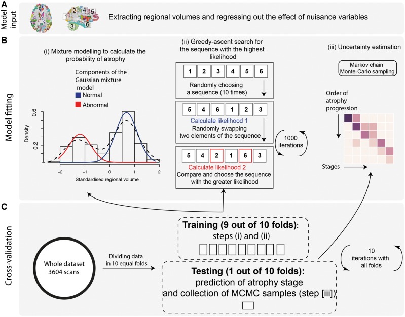
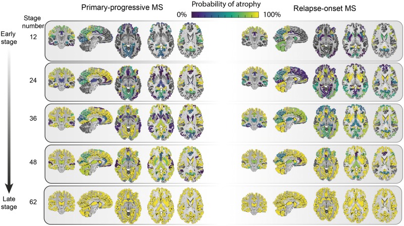
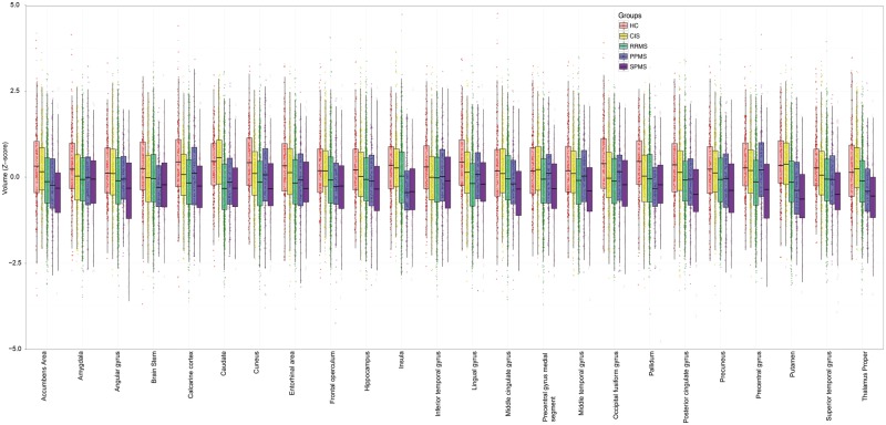
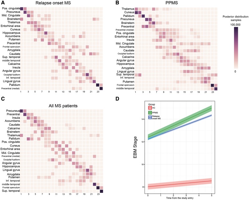
See Stankoff and Louapre (doi:10.1093/brain/awy114) for a scientific commentary on this article.Grey matter atrophy is present from the earliest stages of multiple sclerosis, but its temporal ordering is poorly understood. We aimed to determine the sequence in which grey matter regions become atrophic in multiple sclerosis and its association with disability accumulation. In this longitudinal study, we included 1417 subjects: 253 with clinically isolated syndrome, 708 with relapsing-remitting multiple sclerosis, 128 with secondary-progressive multiple sclerosis, 125 with primary-progressive multiple sclerosis, and 203 healthy control subjects from seven European centres. Subjects underwent repeated MRI (total number of scans 3604); the mean follow-up for patients was 2.41 years (standard deviation = 1.97). Disability was scored using the Expanded Disability Status Scale. We calculated the volume of brain grey matter regions and brainstem using an unbiased within-subject template and used an established data-driven event-based model to determine the sequence of occurrence of atrophy and its uncertainty. We assigned each subject to a specific event-based model stage, based on the number of their atrophic regions. Linear mixed-effects models were used to explore associations between the rate of increase in event-based model stages, and T2 lesion load, disease-modifying treatments, comorbidity, disease duration and disability accumulation. The first regions to become atrophic in patients with clinically isolated syndrome and relapse-onset multiple sclerosis were the posterior cingulate cortex and precuneus, followed by the middle cingulate cortex, brainstem and thalamus. A similar sequence of atrophy was detected in primary-progressive multiple sclerosis with the involvement of the thalamus, cuneus, precuneus, and pallidum, followed by the brainstem and posterior cingulate cortex. The cerebellum, caudate and putamen showed early atrophy in relapse-onset multiple sclerosis and late atrophy in primary-progressive multiple sclerosis. Patients with secondary-progressive multiple sclerosis showed the highest event-based model stage (the highest number of atrophic regions, P < 0.001) at the study entry. All multiple sclerosis phenotypes, but clinically isolated syndrome, showed a faster rate of increase in the event-based model stage than healthy controls. T2 lesion load and disease duration in all patients were associated with increased event-based model stage, but no effects of disease-modifying treatments and comorbidity on event-based model stage were observed. The annualized rate of event-based model stage was associated with the disability accumulation in relapsing-remitting multiple sclerosis, independent of disease duration (P < 0.0001). The data-driven staging of atrophy progression in a large multiple sclerosis sample demonstrates that grey matter atrophy spreads to involve more regions over time. The sequence in which regions become atrophic is reasonably consistent across multiple sclerosis phenotypes. The spread of atrophy was associated with disease duration and with disability accumulation over time in relapsing-remitting multiple sclerosis.

摘要

请参阅 Stankoff 和 Louapre 的科学评论(doi:10.1093/brain/awy114)。灰质萎缩存在于多发性硬化症的早期阶段,但它的时间顺序尚不清楚。我们旨在确定多发性硬化症中灰质区域萎缩的顺序及其与残疾积累的关系。在这项纵向研究中,我们纳入了来自七个欧洲中心的 1417 名受试者:253 名患有临床孤立综合征,708 名患有复发缓解型多发性硬化症,128 名患有继发进展型多发性硬化症,125 名患有原发进展型多发性硬化症,203 名健康对照者。受试者接受了重复的 MRI(共进行了 3604 次扫描);患者的平均随访时间为 2.41 年(标准差=1.97)。残疾使用扩展残疾状态量表进行评分。我们使用无偏的个体内模板计算了脑灰质区域和脑干的体积,并使用既定的数据驱动事件模型来确定萎缩的发生顺序及其不确定性。我们根据患者的萎缩区域数量,将每个患者分配到特定的基于事件的模型阶段。使用线性混合效应模型来探索基于事件的模型阶段增加率与 T2 病变负荷、疾病修正治疗、合并症、疾病持续时间和残疾积累之间的关联。在临床孤立综合征和复发发作型多发性硬化症患者中,首先发生萎缩的区域是后扣带回皮质和楔前叶,其次是中扣带回皮质、脑干和丘脑。原发性进行性多发性硬化症也检测到类似的萎缩顺序,涉及丘脑、楔前叶、楔叶和苍白球,其次是脑干和后扣带回皮质。小脑、尾状核和壳核在复发发作型多发性硬化症中较早发生萎缩,在原发性进行性多发性硬化症中较晚发生萎缩。继发性进行性多发性硬化症患者在研究开始时具有最高的基于事件的模型阶段(最高数量的萎缩区域,P<0.001)。所有多发性硬化症表型,但临床孤立综合征,均显示出比健康对照组更快的基于事件的模型阶段增加率。所有患者的 T2 病变负荷和疾病持续时间均与基于事件的模型阶段增加有关,但未观察到疾病修正治疗和合并症对基于事件的模型阶段的影响。基于事件的模型阶段的年化率与复发缓解型多发性硬化症的残疾积累有关,与疾病持续时间无关(P<0.0001)。在大型多发性硬化症样本中,基于数据驱动的萎缩进展分期表明,灰质萎缩随着时间的推移向更多区域扩散。各多发性硬化症表型之间萎缩区域的发生顺序相当一致。萎缩的扩散与疾病持续时间以及复发缓解型多发性硬化症中随时间推移的残疾积累有关。

https://cdn.ncbi.nlm.nih.gov/pmc/blobs/5a9b/5995197/865babd8ccdf/awy088f4.jpg
https://cdn.ncbi.nlm.nih.gov/pmc/blobs/5a9b/5995197/41c2adefbd6c/awy088f1.jpg
https://cdn.ncbi.nlm.nih.gov/pmc/blobs/5a9b/5995197/fe7d02065028/awy088f2.jpg
https://cdn.ncbi.nlm.nih.gov/pmc/blobs/5a9b/5995197/e26fc55bf9ad/awy088f3.jpg
https://cdn.ncbi.nlm.nih.gov/pmc/blobs/5a9b/5995197/865babd8ccdf/awy088f4.jpg

相似文献

1
Progression of regional grey matter atrophy in multiple sclerosis.多发性硬化症患者的区域性灰质萎缩进展。
Brain. 2018 Jun 1;141(6):1665-1677. doi: 10.1093/brain/awy088.
2
Predicting cognitive decline in multiple sclerosis: a 5-year follow-up study.多发性硬化症认知能力下降的预测:一项 5 年随访研究。
Brain. 2018 Sep 1;141(9):2605-2618. doi: 10.1093/brain/awy202.
3
Brain atrophy and physical disability in primary progressive multiple sclerosis: A volumetric study.原发性进行性多发性硬化症中的脑萎缩与身体残疾:一项容积研究。
Neuroradiol J. 2015 Jun;28(3):354-8. doi: 10.1177/1971400915594984.
4
Cervical cord area is associated with infratentorial grey and white matter volume predominantly in relapsing-remitting multiple sclerosis: A study using semi-automated cord volumetry and voxel-based morphometry.在复发缓解型多发性硬化症中,颈髓区域主要与幕下灰质和白质体积相关:一项使用半自动脊髓容积测量法和基于体素的形态测量学的研究
Mult Scler Relat Disord. 2015 May;4(3):264-72. doi: 10.1016/j.msard.2015.04.003. Epub 2015 Apr 15.
5
Prognostic value of single-subject grey matter networks in early multiple sclerosis.单个体灰质网络对早期多发性硬化症的预后价值。
Brain. 2024 Jan 4;147(1):135-146. doi: 10.1093/brain/awad288.
6
Spatial distribution of multiple sclerosis lesions in the cervical spinal cord.颈髓多发性硬化病灶的空间分布。
Brain. 2019 Mar 1;142(3):633-646. doi: 10.1093/brain/awy352.
7
Sample size requirements for one-year treatment effects using deep gray matter volume from 3T MRI in progressive forms of multiple sclerosis.利用3T磁共振成像(MRI)的深部灰质体积对进展型多发性硬化症一年治疗效果的样本量要求。
Int J Neurosci. 2017 Nov;127(11):971-980. doi: 10.1080/00207454.2017.1283313. Epub 2017 Feb 2.
8
Cortical grey matter sodium accumulation is associated with disability and secondary progressive disease course in relapse-onset multiple sclerosis.皮质灰质钠离子积累与复发缓解型多发性硬化的残疾和继发性进行性疾病过程有关。
J Neurol Neurosurg Psychiatry. 2019 Jul;90(7):755-760. doi: 10.1136/jnnp-2018-319634. Epub 2019 Apr 4.
9
Deep gray matter volume loss drives disability worsening in multiple sclerosis.深部灰质体积损失导致多发性硬化症残疾恶化。
Ann Neurol. 2018 Feb;83(2):210-222. doi: 10.1002/ana.25145. Epub 2018 Feb 6.
10
Cortical lesion load associates with progression of disability in multiple sclerosis.皮质病变负荷与多发性硬化症残疾进展相关。
Brain. 2012 Oct;135(Pt 10):2952-61. doi: 10.1093/brain/aws246.

引用本文的文献

1
Choroid plexus volume in multiple sclerosis: a systematic review and meta-analysis of an emerging imaging biomarker.多发性硬化症中脉络丛体积:对一种新兴成像生物标志物的系统评价和荟萃分析
Eur J Med Res. 2025 Aug 18;30(1):762. doi: 10.1186/s40001-025-03026-z.
2
Gray matter sodium accumulation and atrophy in relapsing-remitting multiple sclerosis correspond to neurotransmitter maps.复发缓解型多发性硬化症中的灰质钠积累和萎缩与神经递质图谱相对应。
Eur Radiol. 2025 Jul 31. doi: 10.1007/s00330-025-11892-8.
3
The Impact of Gray Matter Structural Changes on Clinical Disability in Multiple Sclerosis: Voxel-and Surface-Based Analyses.

本文引用的文献

1
Deep gray matter volume loss drives disability worsening in multiple sclerosis.深部灰质体积损失导致多发性硬化症残疾恶化。
Ann Neurol. 2018 Feb;83(2):210-222. doi: 10.1002/ana.25145. Epub 2018 Feb 6.
2
Progression of regional atrophy in the left hemisphere contributes to clinical and cognitive deterioration in multiple sclerosis: A 5-year study.左侧半球区域性萎缩的进展导致多发性硬化症的临床和认知恶化:一项 5 年研究。
Hum Brain Mapp. 2017 Nov;38(11):5648-5665. doi: 10.1002/hbm.23755. Epub 2017 Aug 9.
3
Age-related small vessel disease: a potential contributor to neurodegeneration in multiple sclerosis.
灰质结构变化对多发性硬化症临床残疾的影响:基于体素和表面的分析
Neuroinformatics. 2025 Jul 23;23(3):39. doi: 10.1007/s12021-025-09740-7.
4
Thalamic Network Controllability Predicts Cognitive Impairment in Multiple Sclerosis.丘脑网络可控性可预测多发性硬化症中的认知障碍。
Hum Brain Mapp. 2025 Jul;46(10):e70284. doi: 10.1002/hbm.70284.
5
Neuroimaging phenotype characterization of early aggressive and late severe multiple sclerosis: a case-control study.早期侵袭性和晚期重症多发性硬化的神经影像学表型特征:一项病例对照研究。
Brain Commun. 2025 Jun 24;7(4):fcaf254. doi: 10.1093/braincomms/fcaf254. eCollection 2025.
6
Accelerated frontal grey matter atrophy in relapsing remitting multiple sclerosis.复发缓解型多发性硬化症患者额叶灰质加速萎缩。
medRxiv. 2025 Jul 2:2025.07.01.25330635. doi: 10.1101/2025.07.01.25330635.
7
Follicle on the Roof: Tertiary Lymphoid Structures in Central Nervous System Autoimmunity.位于脑顶部的滤泡:中枢神经系统自身免疫中的三级淋巴结构
Immunol Rev. 2025 Jul;332(1):e70045. doi: 10.1111/imr.70045.
8
Decreased mitochondrial activity in the demyelinating cerebellum of progressive multiple sclerosis and chronic EAE contributes to Purkinje cell loss.进行性多发性硬化症和慢性实验性自身免疫性脑脊髓炎脱髓鞘小脑中线粒体活性降低导致浦肯野细胞丢失。
Proc Natl Acad Sci U S A. 2025 Jun 24;122(25):e2421806122. doi: 10.1073/pnas.2421806122. Epub 2025 Jun 16.
9
Assessing the role of volumetric brain information in multiple sclerosis progression.评估脑容量信息在多发性硬化症进展中的作用。
Comput Struct Biotechnol J. 2025 May 12;27:2014-2033. doi: 10.1016/j.csbj.2025.05.003. eCollection 2025.
10
Advances in Duchenne Muscular Dystrophy: Diagnostic Techniques and Dystrophin Domain Insights.杜兴氏肌肉营养不良症的进展:诊断技术与肌营养不良蛋白结构域见解
Int J Mol Sci. 2025 Apr 10;26(8):3579. doi: 10.3390/ijms26083579.
年龄相关性小血管疾病:多发性硬化症神经退行性变的一个潜在因素。
Brain Pathol. 2017 Nov;27(6):707-722. doi: 10.1111/bpa.12460. Epub 2017 Mar 12.
4
Relationship of grey and white matter abnormalities with distance from the surface of the brain in multiple sclerosis.多发性硬化症患者脑表面距离与灰白质异常的关系。
J Neurol Neurosurg Psychiatry. 2016 Nov;87(11):1212-1217. doi: 10.1136/jnnp-2016-313979. Epub 2016 Sep 6.
5
The topograpy of demyelination and neurodegeneration in the multiple sclerosis brain.多发性硬化症脑部脱髓鞘和神经变性的拓扑结构。
Brain. 2016 Mar;139(Pt 3):807-15. doi: 10.1093/brain/awv398. Epub 2016 Feb 8.
6
White and gray matter damage in primary progressive MS: The chicken or the egg?原发性进展型多发性硬化症中的白质和灰质损伤:先有鸡还是先有蛋?
Neurology. 2016 Jan 12;86(2):170-6. doi: 10.1212/WNL.0000000000002237. Epub 2015 Dec 16.
7
Cortical atrophy patterns in multiple sclerosis are non-random and clinically relevant.多发性硬化症中的皮质萎缩模式具有非随机性且与临床相关。
Brain. 2016 Jan;139(Pt 1):115-26. doi: 10.1093/brain/awv337. Epub 2015 Dec 4.
8
Association of Deep Gray Matter Damage With Cortical and Spinal Cord Degeneration in Primary Progressive Multiple Sclerosis.原发性进行性多发性硬化症中深部灰质损伤与皮质和脊髓退化的关联。
JAMA Neurol. 2015 Dec;72(12):1466-74. doi: 10.1001/jamaneurol.2015.1897.
9
Regional Distribution and Evolution of Gray Matter Damage in Different Populations of Multiple Sclerosis Patients.多发性硬化症患者不同群体中灰质损伤的区域分布与演变
PLoS One. 2015 Aug 12;10(8):e0135428. doi: 10.1371/journal.pone.0135428. eCollection 2015.
10
The brain's default mode network.大脑的默认模式网络。
Annu Rev Neurosci. 2015 Jul 8;38:433-47. doi: 10.1146/annurev-neuro-071013-014030. Epub 2015 May 4.