Department of Medicine, Cardiovascular Medicine Unit, Center for Molecular Medicine, Karolinska Institute, Karolinska University Hospital, Stockholm, Sweden.
Department of Medicine, University of Valencia, Valencia, Spain.
Oxid Med Cell Longev. 2018 Mar 18;2018:1975167. doi: 10.1155/2018/1975167. eCollection 2018.
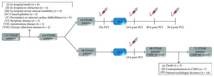
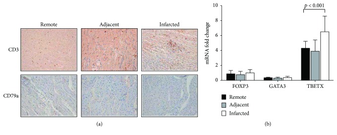
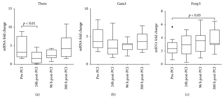
ST-segment elevation myocardial infarction (STEMI) is the most severe outcome of coronary artery disease. Despite rapid reperfusion of the artery, acute irrigation of the cardiac tissue is associated with increased inflammation. While innate immune response in STEMI is well described, an in-depth characterization of adaptive immune cell dynamics and their potential role remains elusive. We performed a translational study using a controlled porcine reperfusion model of STEMI and the analysis of lymphocyte subsets in 116 STEMI patients undergoing percutaneous coronary intervention (PCI). In the animal model, a sharp drop in circulating T lymphocytes occurred within the first hours after reperfusion. Notably, increased apoptosis of circulating lymphocytes and infiltration of proinflammatory Th1 lymphocytes in the heart were observed 48 h after reperfusion. Similarly, in STEMI patients, a sharp drop in circulating T lymphocyte subsets occurred within the first 24 h post-PCI. A cardiac magnetic resonance (CMR) evaluation of these patients revealed an inverse association between 24 h circulating T lymphocyte numbers and infarction size at 1-week and 6-month post-PCI. Our translational approach revealed striking changes in the circulating and tissue-infiltrating T lymphocyte repertoire in response to ischemia-reperfusion. These findings may help in developing new diagnostic and therapeutic approaches for coronary diseases.
ST 段抬高型心肌梗死(STEMI)是冠状动脉疾病最严重的后果。尽管动脉迅速再灌注,但急性心肌组织灌流与炎症增加有关。虽然 STEMI 的固有免疫反应已有很好的描述,但适应性免疫细胞动力学及其潜在作用的深入特征仍难以捉摸。我们使用 STEMI 的受控猪再灌注模型和 116 例行经皮冠状动脉介入治疗(PCI)的 STEMI 患者的淋巴细胞亚群分析进行了一项转化研究。在动物模型中,再灌注后最初几小时内循环 T 淋巴细胞数量急剧下降。值得注意的是,再灌注后 48 小时观察到循环淋巴细胞凋亡增加和促炎 Th1 淋巴细胞浸润心脏。同样,在 STEMI 患者中,PCI 后 24 小时内循环 T 淋巴细胞亚群数量急剧下降。对这些患者的心脏磁共振(CMR)评估显示,PCI 后 1 周和 6 个月时 24 小时循环 T 淋巴细胞数量与梗死面积呈负相关。我们的转化方法揭示了对缺血再灌注的循环和组织浸润 T 淋巴细胞库的惊人变化。这些发现可能有助于为冠状动脉疾病开发新的诊断和治疗方法。