Williams James C, Zarse Chad A, Jackson Molly E, Lingeman James E, McAteer James A
Department of Anatomy and Cell Biology, Indiana University School of Medicine, Indianapolis, IN 46202, USA.
Methodist Hospital Institute for Kidney Stone Disease, 1801 North Senate Boulevard, Suite 220, Indianapolis, Indiana 46202. USA.
AIP Conf Proc. 2007;900:326-339. doi: 10.1063/1.2723592. Epub 2007 Apr 5.
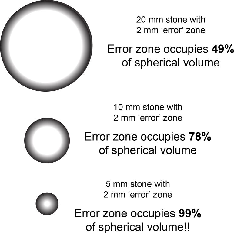
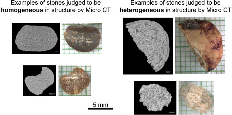
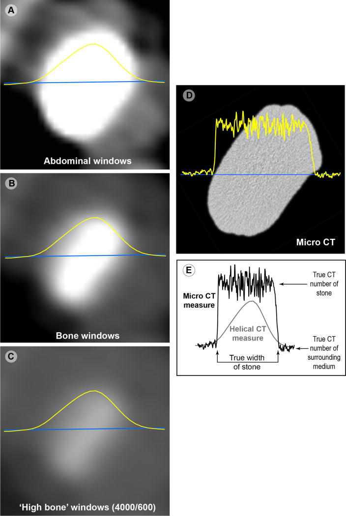
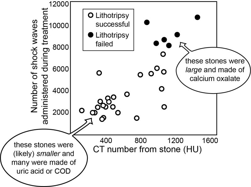
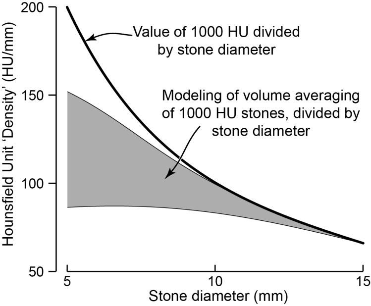
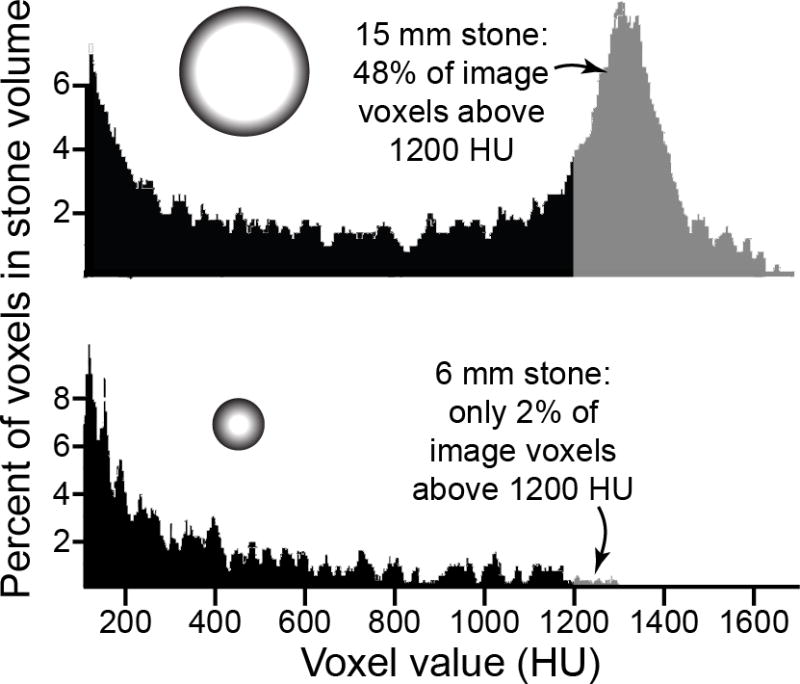
Great variability exists in the response of urinary stones to SWL, and this is true even for stones composed of the same mineral. Efforts have been made to predict stone fragility to shock waves using computed tomography (CT) patient images, but most work to date has focused on the use of stone CT number (i.e., Hounsfield units). This is an easy number to measure on a patient stone, but its value depends on a number of factors, including the relationship of the size of the stone to the resolution (i.e., the slicewidth) of the CT scan. Studies that have shown a relationship between stone CT number and failure in SWL are reviewed, and all are shown to suffer from error due to stone size, which was not accounted for in the use of Hounsfield unit values. Preliminary data are then presented for a study of calcium oxalate monohydrate (COM) stones, in which stone structure-rather than simple CT number values-is shown to correlate with fragility to shock waves. COM stones that were observed to have structure by micro CT (e.g., voids, apatite regions, unusual shapes) broke to completion in about half the number of shock waves required for COM stones that were observed to be homogeneous in structure by CT. This result suggests another direction for the use of CT in predicting success of SWL: the use of CT to view stone structure, rather than simply measuring stone CT number. Viewing stone structure by CT requires the use of different viewing windows than those typically used for examining patient scans, but much research to date indicates that stone structure can be observed in the clinical setting. Future clinical studies will need to be done to verify the relationship between stone structure observed by CT and stone fragility in SWL.
尿路结石对体外冲击波碎石术(SWL)的反应存在很大差异,即使是由相同矿物质组成的结石也是如此。人们已努力利用计算机断层扫描(CT)患者图像来预测结石对冲击波的易碎性,但迄今为止,大多数工作都集中在使用结石CT值(即亨氏单位)上。这是一个在患者结石上易于测量的数值,但其值取决于许多因素,包括结石大小与CT扫描分辨率(即层厚)的关系。本文回顾了那些显示结石CT值与SWL治疗失败之间存在关联的研究,结果表明,所有这些研究都因结石大小而存在误差,而在使用亨氏单位值时并未考虑这一因素。随后给出了一项关于一水合草酸钙(COM)结石研究的初步数据,其中显示结石结构而非简单的CT值与对冲击波的易碎性相关。通过显微CT观察到具有结构的COM结石(如孔隙、磷灰石区域、异常形状),其完全破碎所需的冲击波次数约为通过CT观察到结构均匀的COM结石的一半。这一结果为利用CT预测SWL治疗成功率指明了另一个方向:利用CT观察结石结构,而不仅仅是测量结石CT值。通过CT观察结石结构需要使用与通常用于检查患者扫描图像不同的观察窗,但迄今为止的大量研究表明,在临床环境中可以观察到结石结构。未来需要开展临床研究来验证CT观察到的结石结构与SWL中结石易碎性之间的关系。