Zhang Yi, Zhao Lin, Wang Lijun, Yang Xiting, Zhou Aiyi, Wang Jianming
Department of Ophthalmology, the Second Affiliated Hospital of Xi'an Jiaotong University.
Mol Vis. 2018 Apr 26;24:340-352. eCollection 2018.
To investigate the role of placental growth factor (PGF) in the epithelial-mesenchymal transition (EMT) of ARPE-19 cells under hypoxia, and whether the NF-κB signaling pathway is involved in this process.
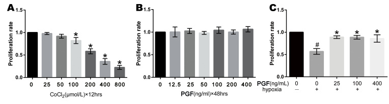
ARPE-19 cells were treated in five groups: a control group, hypoxia group, PGF group, hypoxia+PGF group, and NF-κB-blocked group. A chemical hypoxia model was established in the ARPE-19 cells by adding CoCl to the culture medium. The morphological changes after treatment were observed. The proliferation rates were measured with 3-(4,5-dimethyl-2-thiazolyl)-2,5-diphenyl-2H-tetrazolium bromide (MTT) assay. The migration abilities were measured with scratch assay. The EMT biomarkers were measured with quantitative real-time PCR (qRT-PCR), western blotting, and immunofluorescence. The relative protein expression of components of the NF-κB signaling pathway was measured with western blotting and immunofluorescence.
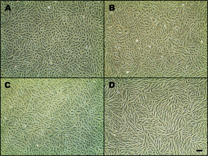
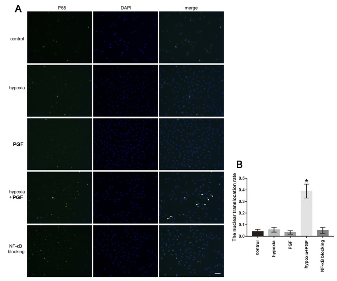
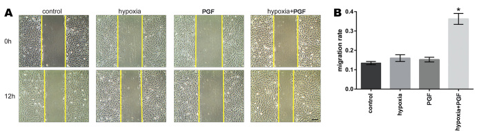
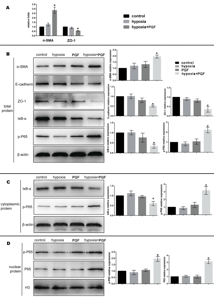
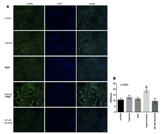
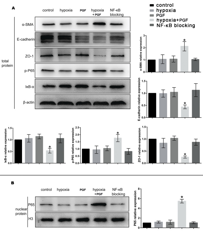
Cells treated with PGF under hypoxia exhibited morphological changes consistent with the transition from an epithelial to a mesenchymal phenotype. In the ARPE-19 cells, exogenous PGF under hypoxia increased the proliferation rate compared to the rate under hypoxia alone (p<0.05) and increased the migration rate (p<0.05). Treatment of hypoxia-exposed cells with PGF caused decreased expression of the epithelial biomarkers E-cadherin and ZO-1 (both p<0.05) and increased expression of the mesenchymal marker α-SMA (p<0.05) by enhancing the phosphorylation of NF-κB p65 of the total protein, promoting the translocation of p65 to the nucleus, and inducing the degradation of IκB-α (a negative regulator of the NF-κB pathway) in the ARPE-19 cells. Additionally, the effect of PGF-induced EMT in the ARPE-19 cells under hypoxia was counteracted with BAY 11-7082 (a selective NF-κB inhibitor).
Exogenous PGF promotes EMT-like changes in ARPE-19 cells under hypoxia by activating the NF-κB signaling pathway. The study results suggest that PGF may play a role in scar formation in neovascular age-related macular degeneration (AMD) and that the inhibition of PGF may be a promising target for the prevention and treatment of AMD.
探讨胎盘生长因子(PGF)在缺氧条件下对ARPE - 19细胞上皮 - 间充质转化(EMT)中的作用,以及核因子κB(NF - κB)信号通路是否参与此过程。
将ARPE - 19细胞分为五组:对照组、缺氧组、PGF组、缺氧 + PGF组和NF - κB阻断组。通过向培养基中添加氯化钴(CoCl)在ARPE - 19细胞中建立化学缺氧模型。观察处理后的形态学变化。用3 -(4,5 - 二甲基 - 2 - 噻唑基)- 2,5 - 二苯基 - 2H - 四氮唑溴盐(MTT)法测定增殖率。用划痕试验测定迁移能力。用定量实时聚合酶链反应(qRT - PCR)、蛋白质免疫印迹法和免疫荧光法检测EMT生物标志物。用蛋白质免疫印迹法和免疫荧光法测定NF - κB信号通路各组分的相对蛋白表达。
缺氧条件下用PGF处理的细胞表现出与上皮细胞向间充质细胞表型转变一致的形态学变化。在ARPE - 19细胞中,缺氧条件下外源性PGF与单纯缺氧相比增加了增殖率(p < 0.05)并提高了迁移率(p < 0.05)。用PGF处理缺氧暴露的细胞导致上皮生物标志物E - 钙黏蛋白和紧密连接蛋白1(ZO - 1)的表达降低(均p < 0.05),并通过增强ARPE - 19细胞中总蛋白NF - κB p65的磷酸化、促进p65向细胞核的转位以及诱导IκB - α(NF - κB通路的负调节因子)的降解,使间充质标志物α - 平滑肌肌动蛋白(α - SMA)的表达增加(p < 0.05)。此外,BAY 11 - 7082(一种选择性NF - κB抑制剂)抵消了缺氧条件下PGF诱导ARPE - 19细胞发生EMT的作用。
外源性PGF通过激活NF - κB信号通路促进缺氧条件下ARPE - 19细胞发生类似EMT的变化。研究结果表明,PGF可能在新生血管性年龄相关性黄斑变性(AMD)的瘢痕形成中起作用,抑制PGF可能是预防和治疗AMD的一个有前景的靶点。