Pavon Lorena Favaro, Sibov Tatiana Tais, Caminada de Toledo Silvia Regina, Mara de Oliveira Daniela, Cabral Francisco Romero, Gabriel de Souza Jean, Boufleur Pamela, Marti Luciana C, Malheiros Jackeline Moraes, Ferreira da Cruz Edgar, Paiva Fernando F, Malheiros Suzana M F, de Paiva Neto Manoel A, Tannús Alberto, Mascarenhas de Oliveira Sérgio, Silva Nasjla Saba, Cappellano Andrea Maria, Petrilli Antonio Sérgio, Chudzinski-Tavassi Ana Marisa, Cavalheiro Sérgio
Department of Neurology and Neurosurgery, Escola Paulista de Medicina (EPM), Universidade Federal de São Paulo (UNIFESP), São Paulo, Brazil.
Pediatric Oncology Institute, Grupo de Apoio ao Adolescente e à Criança com Câncer (GRAACC), Escola Paulista de Medicina (EPM), Universidade Federal de São Paulo (UNIFESP), São Paulo, Brazil.
Oncotarget. 2018 Apr 24;9(31):21731-21743. doi: 10.18632/oncotarget.24932.
Ependymoma (EPN), the third most common pediatric brain tumor, is a central nervous system (CNS) malignancy originating from the walls of the ventricular system. Surgical resection followed by radiation therapy has been the primary treatment for most pediatric intracranial EPNs. Despite numerous studies into the prognostic value of histological classification, the extent of surgical resection and adjuvant radiotherapy, there have been relatively few studies into the molecular and cellular biology of EPNs.
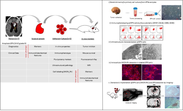
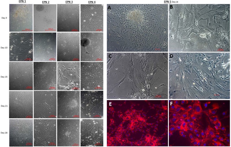
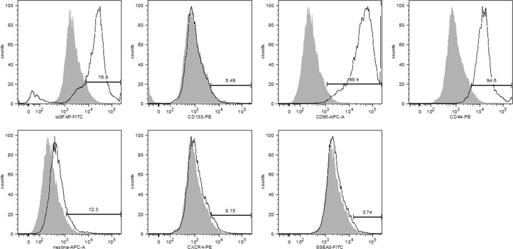
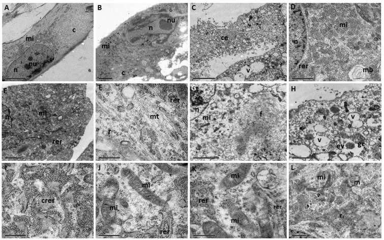
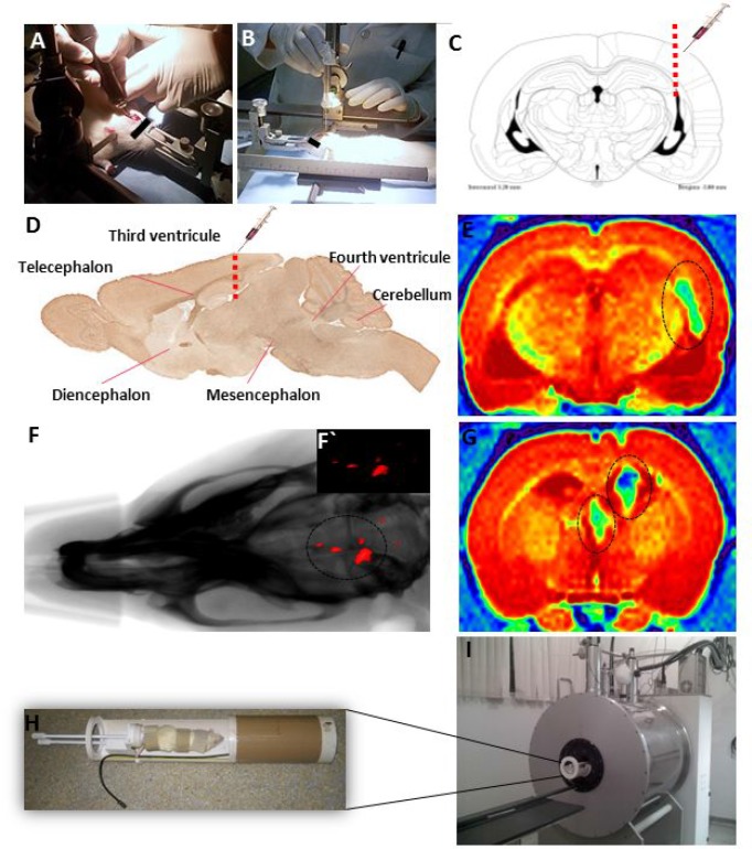
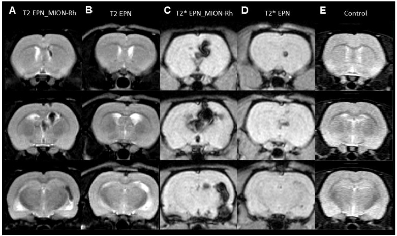
We elucidated the ultrastructure of the cultured EPN cells and characterized their profile of immunophenotypic pluripotency markers (CD133, CD90, SSEA-3, CXCR4). We established an experimental EPN model by the intracerebroventricular infusion of EPN cells labeled with multimodal iron oxide nanoparticles (MION), thereby generating a tumor and providing a clinically relevant animal model. MRI analysis was shown to be a valuable tool when combined with effective MION labeling techniques to accompany EPN growth.
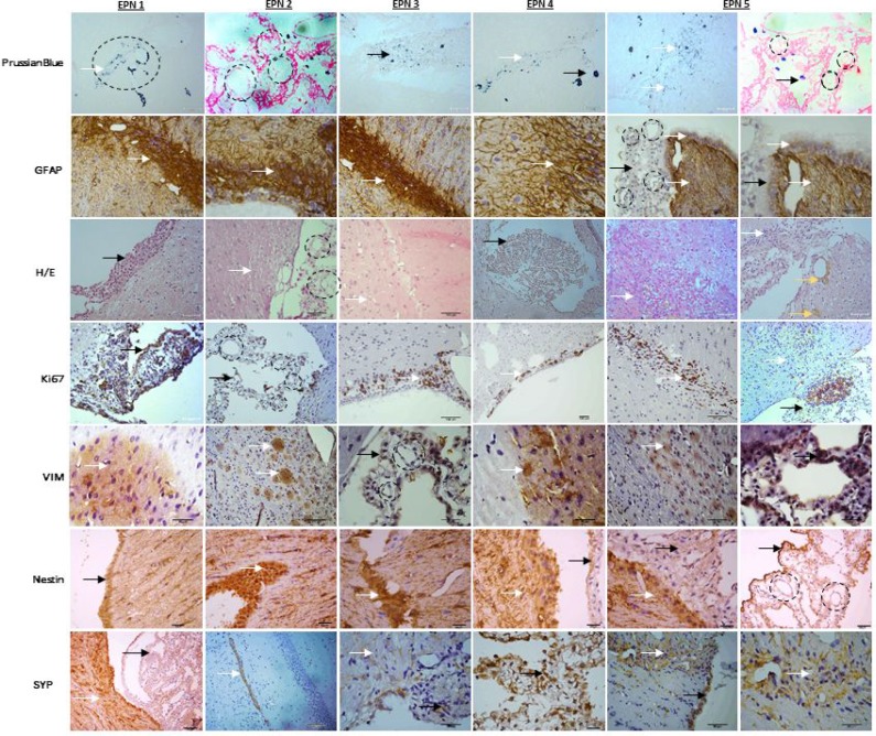
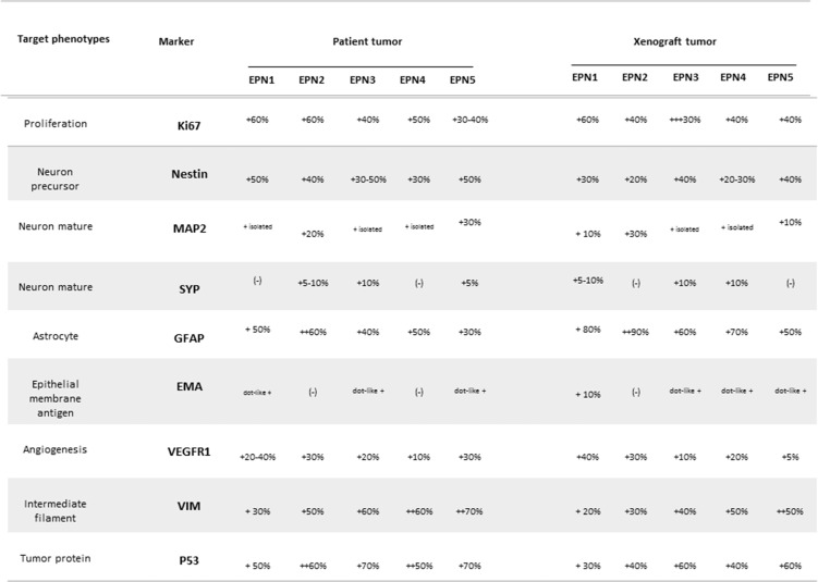
We demonstrated that GFAP/CD133+CD90+/CD44+ EPN cells maintained key histopathological and growth characteristics of the original patient tumor. The characterization of EPN cells and the experimental model could facilitate biological studies and preclinical drug screening for pediatric EPNs.
In this work, we established notoriously challenging primary cell culture of anaplastic EPNs (WHO grade III) localized in the posterior fossa (PF), using EPNs obtained from 1 to 10-year-old patients ( = 07), and then characterized their immunophenotype and ultrastructure to finally develop a xenograft model.
室管膜瘤(EPN)是儿童第三常见的脑肿瘤,是一种起源于脑室系统壁的中枢神经系统(CNS)恶性肿瘤。手术切除后进行放射治疗一直是大多数儿童颅内EPN的主要治疗方法。尽管对组织学分类、手术切除范围和辅助放疗的预后价值进行了大量研究,但对EPN的分子和细胞生物学研究相对较少。
我们阐明了培养的EPN细胞的超微结构,并对其免疫表型多能性标志物(CD133、CD90、SSEA-3、CXCR4)的特征进行了描述。我们通过脑室内注入用多模态氧化铁纳米颗粒(MION)标记的EPN细胞建立了一个实验性EPN模型,从而产生肿瘤并提供一个临床相关的动物模型。当与有效的MION标记技术相结合以伴随EPN生长时,MRI分析被证明是一种有价值的工具。
我们证明了GFAP/CD133+CD90+/CD44+ EPN细胞保留了原始患者肿瘤的关键组织病理学和生长特征。EPN细胞的特征描述和实验模型有助于儿童EPN的生物学研究和临床前药物筛选。
在这项工作中,我们使用从1至10岁患者(n = 07)获得的EPN,建立了位于后颅窝(PF)的间变性EPN(世界卫生组织III级)极具挑战性的原代细胞培养,然后对其免疫表型和超微结构进行了表征,最终建立了一个异种移植模型。