• 文献检索
  • 文档翻译
  • 深度研究
  • 学术资讯
  • Suppr Zotero 插件Zotero 插件
  • 邀请有礼
  • 套餐&价格
  • 历史记录
应用&插件
Suppr Zotero 插件Zotero 插件浏览器插件Mac 客户端Windows 客户端微信小程序
定价
高级版会员购买积分包购买API积分包
服务
文献检索文档翻译深度研究API 文档MCP 服务
关于我们
关于 Suppr公司介绍联系我们用户协议隐私条款
关注我们

Suppr 超能文献

核心技术专利:CN118964589B侵权必究
粤ICP备2023148730 号-1Suppr @ 2026

文献检索

告别复杂PubMed语法,用中文像聊天一样搜索,搜遍4000万医学文献。AI智能推荐,让科研检索更轻松。

立即免费搜索

文件翻译

保留排版,准确专业,支持PDF/Word/PPT等文件格式,支持 12+语言互译。

免费翻译文档

深度研究

AI帮你快速写综述,25分钟生成高质量综述,智能提取关键信息,辅助科研写作。

立即免费体验

轻度创伤性脑损伤的混合扩散成像。

Hybrid Diffusion Imaging in Mild Traumatic Brain Injury.

机构信息

1 Department of Radiology and Imaging Sciences, Indiana University School of Medicine , Indianapolis, Indiana.

2 Department of Epidemiology and Biostatistics, School of Public Health, Indiana University , Bloomington, Indiana.

出版信息

J Neurotrauma. 2018 Oct 15;35(20):2377-2390. doi: 10.1089/neu.2017.5566. Epub 2018 Jul 6.

DOI:10.1089/neu.2017.5566
PMID:29786463
原文链接:https://pmc.ncbi.nlm.nih.gov/articles/PMC6196746/
Abstract

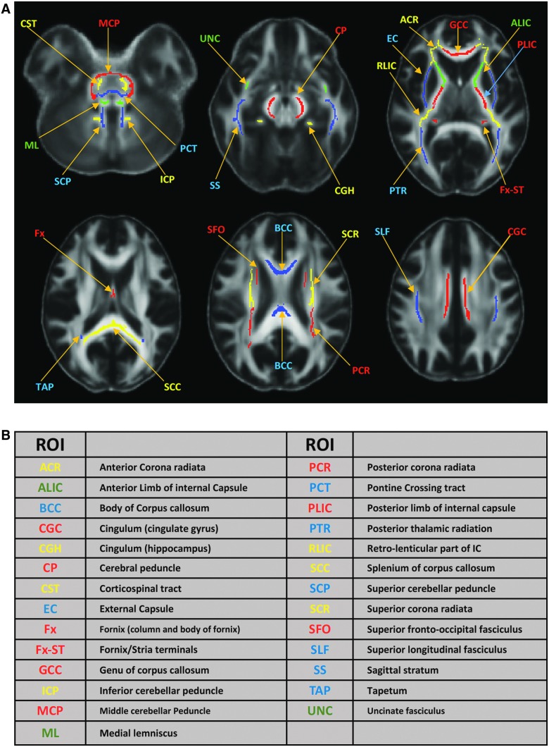
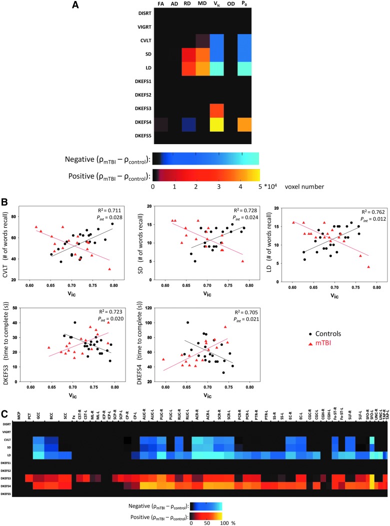
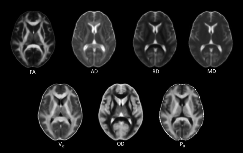
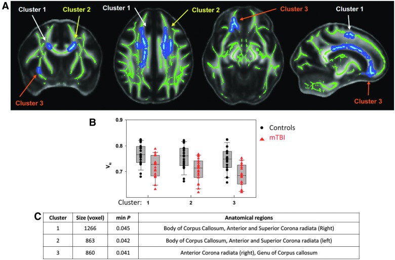
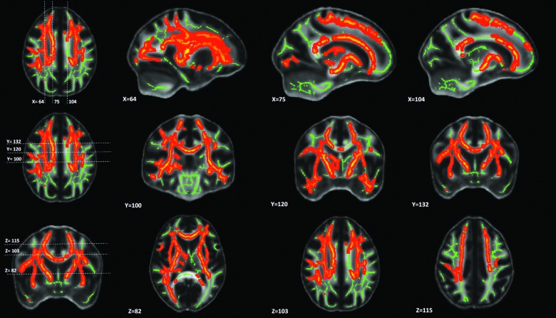
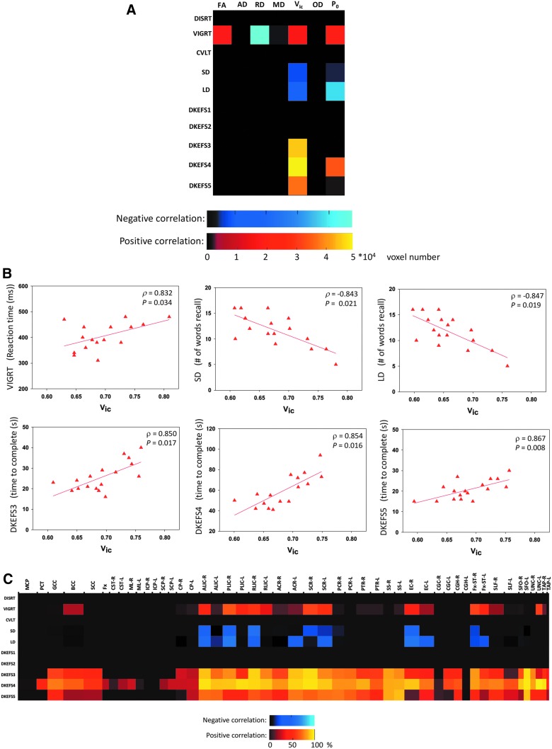
Mild traumatic brain injury (mTBI) is an important public health problem. Although conventional medical imaging techniques can detect moderate-to-severe injuries, they are relatively insensitive to mTBI. In this study, we used hybrid diffusion imaging (HYDI) to detect white matter alterations in 19 patients with mTBI and 23 other trauma control patients. Within 15 days (standard deviation = 10) of brain injury, all subjects underwent magnetic resonance HYDI and were assessed with a battery of neuropsychological tests of sustained attention, memory, and executive function. Tract-based spatial statistics (TBSS) was used for voxel-wise statistical analyses within the white matter skeleton to study between-group differences in diffusion metrics, within-group correlations between diffusion metrics and clinical outcomes, and between-group interaction effects. The advanced diffusion imaging techniques, including neurite orientation dispersion and density imaging (NODDI) and q-space analyses, appeared to be more sensitive then classic diffusion tensor imaging. Only NODDI-derived intra-axonal volume fraction (V) demonstrated significant group differences (i.e., 5-9% lower in the injured brain). Within the mTBI group, V and a q-space measure, P, correlated with 6 of 10 neuropsychological tests, including measures of attention, memory, and executive function. In addition, the direction of correlations differed significantly between groups (R > 0.71 and p < 0.03). Specifically, in the control group, higher V and P were associated with better performances on clinical assessments, whereas in the mTBI group, higher V and P were associated with worse performances with correlation coefficients >0.83. In summary, the NODDI-derived axonal density index and q-space measure for tissue restriction demonstrated superior sensitivity to white matter changes shortly after mTBI. These techniques hold promise as a neuroimaging biomarker for mTBI.

摘要

轻度创伤性脑损伤(mTBI)是一个重要的公共卫生问题。尽管传统的医学成像技术可以检测到中度至重度损伤,但它们对 mTBI 相对不敏感。在这项研究中,我们使用混合扩散成像(HYDI)来检测 19 名 mTBI 患者和 23 名其他创伤对照患者的白质改变。在脑损伤后 15 天内(标准差=10),所有受试者都接受了磁共振 HYDI 检查,并进行了一系列持续注意力、记忆和执行功能的神经心理学测试。基于体素的空间统计学(TBSS)用于白质骨架内的体素统计分析,以研究扩散指标的组间差异、组内扩散指标与临床结果的相关性以及组间的相互作用效应。先进的扩散成像技术,包括神经丝取向分散和密度成像(NODDI)和 q 空间分析,似乎比经典的扩散张量成像更敏感。只有 NODDI 衍生的轴内体积分数(V)表现出显著的组间差异(即损伤大脑中低 5-9%)。在 mTBI 组中,V 和 q 空间测量 P 与 10 项神经心理学测试中的 6 项相关,包括注意力、记忆和执行功能的测试。此外,两组之间的相关性方向差异显著(R>0.71,p<0.03)。具体而言,在对照组中,较高的 V 和 P 与临床评估中的更好表现相关,而在 mTBI 组中,较高的 V 和 P 与更差的表现相关,相关系数>0.83。总之,NODDI 衍生的轴突密度指数和 q 空间测量组织限制显示出对 mTBI 后不久白质变化的更高敏感性。这些技术有望成为 mTBI 的神经影像学生物标志物。

https://cdn.ncbi.nlm.nih.gov/pmc/blobs/cc80/6196746/cafa80bd5747/fig-6.jpg
https://cdn.ncbi.nlm.nih.gov/pmc/blobs/cc80/6196746/59e3abed3d68/fig-1.jpg
https://cdn.ncbi.nlm.nih.gov/pmc/blobs/cc80/6196746/8780b292fc54/fig-2.jpg
https://cdn.ncbi.nlm.nih.gov/pmc/blobs/cc80/6196746/fde5986d8524/fig-3.jpg
https://cdn.ncbi.nlm.nih.gov/pmc/blobs/cc80/6196746/847a7a036a49/fig-4.jpg
https://cdn.ncbi.nlm.nih.gov/pmc/blobs/cc80/6196746/6a1fff81dd1b/fig-5.jpg
https://cdn.ncbi.nlm.nih.gov/pmc/blobs/cc80/6196746/cafa80bd5747/fig-6.jpg

相似文献

1
Hybrid Diffusion Imaging in Mild Traumatic Brain Injury.轻度创伤性脑损伤的混合扩散成像。
J Neurotrauma. 2018 Oct 15;35(20):2377-2390. doi: 10.1089/neu.2017.5566. Epub 2018 Jul 6.
2
Age effects and sex differences in human brain white matter of young to middle-aged adults: A DTI, NODDI, and q-space study.青年至中年成年人脑白质的年龄效应和性别差异:一项扩散张量成像(DTI)、神经突方向离散与密度成像(NODDI)和q空间研究
Neuroimage. 2016 Mar;128:180-192. doi: 10.1016/j.neuroimage.2015.12.033. Epub 2015 Dec 24.
3
Hybrid diffusion imaging reveals altered white matter tract integrity and associations with symptoms and cognitive dysfunction in chronic traumatic brain injury.混合扩散成像揭示了慢性创伤性脑损伤中白质束完整性的改变,并与症状和认知功能障碍有关。
Neuroimage Clin. 2021;30:102681. doi: 10.1016/j.nicl.2021.102681. Epub 2021 Apr 21.
4
Examining brain white matter after pediatric mild traumatic brain injury using neurite orientation dispersion and density imaging: An A-CAP study.应用神经丝取向分散和密度成像技术研究儿科轻度创伤性脑损伤后脑白质:一项 A-CAP 研究。
Neuroimage Clin. 2021;32:102887. doi: 10.1016/j.nicl.2021.102887. Epub 2021 Nov 19.
5
Diffusion Tensor and Kurtosis Imaging Findings the First Year following Mild Traumatic Brain Injury.弥散张量和峰度成像研究在轻度创伤性脑损伤后的第一年发现。
J Neurotrauma. 2023 Mar;40(5-6):457-471. doi: 10.1089/neu.2022.0206. Epub 2022 Dec 9.
6
Evidence of Ongoing Cerebral Microstructural Reorganization in Children With Persisting Symptoms Following Mild Traumatic Brain Injury: A NODDI DTI Analysis.轻度创伤性脑损伤后持续症状儿童的脑微结构重塑的证据:一种 NODDI DTI 分析。
J Neurotrauma. 2024 Jan;41(1-2):41-58. doi: 10.1089/neu.2023.0196. Epub 2023 Nov 29.
7
White matter microstructure in athletes with a history of concussion: Comparing diffusion tensor imaging (DTI) and neurite orientation dispersion and density imaging (NODDI).有脑震荡病史的运动员的白质微观结构:比较扩散张量成像(DTI)和神经突方向离散度与密度成像(NODDI)。
Hum Brain Mapp. 2017 Aug;38(8):4201-4211. doi: 10.1002/hbm.23658. Epub 2017 May 29.
8
High angular resolution diffusion-weighted imaging in mild traumatic brain injury.轻度创伤性脑损伤的高角分辨率扩散加权成像
Neuroimage Clin. 2016 Nov 17;13:174-180. doi: 10.1016/j.nicl.2016.11.016. eCollection 2017.
9
Investigating White Matter Tract Microstructural Changes at Six-Twelve Weeks following Mild Traumatic Brain Injury: A Combined Diffusion Tensor Imaging and Neurite Orientation Dispersion and Density Imaging Study.研究轻度创伤性脑损伤后 6-12 周的白质束微观结构变化:弥散张量成像与神经丝取向弥散和密度成像的联合研究。
J Neurotrauma. 2021 Aug 15;38(16):2255-2263. doi: 10.1089/neu.2020.7310. Epub 2021 Feb 4.
10
Application of neurite orientation dispersion and density imaging to characterize brain microstructural abnormalities in type-2 diabetics with mild cognitive impairment.神经突方向分散和密度成像在 2 型糖尿病伴轻度认知障碍患者脑微结构异常中的应用。
J Magn Reson Imaging. 2019 Sep;50(3):889-898. doi: 10.1002/jmri.26687. Epub 2019 Feb 19.

引用本文的文献

1
Assessment of white matter microstructure integrity in subacute postconcussive vestibular dysfunction using NODDI.使用神经突方向离散度与密度成像(NODDI)评估亚急性脑震荡后前庭功能障碍中的白质微观结构完整性。
Imaging Neurosci (Camb). 2024;2. doi: 10.1162/imag_a_00147. Epub 2024 Apr 10.
2
Significance of the corpus callosum and inferior fronto-occipital fasciculus in recovery after traumatic brain injury.胼胝体和额枕下束在创伤性脑损伤后恢复中的意义。
Neuroradiology. 2025 Mar 11. doi: 10.1007/s00234-025-03577-2.
3
White Matter Alterations in Military Service Members With Remote Mild Traumatic Brain Injury.

本文引用的文献

1
Neurite dispersion: a new marker of multiple sclerosis spinal cord pathology?神经突分散:多发性硬化症脊髓病理学的一个新标志物?
Ann Clin Transl Neurol. 2017 Aug 15;4(9):663-679. doi: 10.1002/acn3.445. eCollection 2017 Sep.
2
White matter microstructure in athletes with a history of concussion: Comparing diffusion tensor imaging (DTI) and neurite orientation dispersion and density imaging (NODDI).有脑震荡病史的运动员的白质微观结构:比较扩散张量成像(DTI)和神经突方向离散度与密度成像(NODDI)。
Hum Brain Mapp. 2017 Aug;38(8):4201-4211. doi: 10.1002/hbm.23658. Epub 2017 May 29.
3
Free water elimination improves test-retest reproducibility of diffusion tensor imaging indices in the brain: A longitudinal multisite study of healthy elderly subjects.
患有慢性轻度创伤性脑损伤的退伍军人的脑白质改变。
JAMA Netw Open. 2024 Apr 1;7(4):e248121. doi: 10.1001/jamanetworkopen.2024.8121.
4
Neurite Damage in Patients with Migraine.偏头痛患者的神经突损伤
Neurol Int. 2024 Feb 29;16(2):299-311. doi: 10.3390/neurolint16020021.
5
Associations of prior concussion severity with brain microstructure using mean apparent propagator magnetic resonance imaging.使用平均表观扩散系数磁共振成像评估既往脑震荡严重程度与脑微观结构的相关性。
Hum Brain Mapp. 2024 Jan;45(1):e26556. doi: 10.1002/hbm.26556. Epub 2023 Dec 29.
6
The relationship between cognition and white matter tract damage after mild traumatic brain injury in a premorbidly healthy, hospitalised adult cohort during the post-acute period.急性后期,病前健康的住院成年队列中轻度创伤性脑损伤后认知与白质束损伤之间的关系。
Front Neurol. 2023 Oct 23;14:1278908. doi: 10.3389/fneur.2023.1278908. eCollection 2023.
7
Machine learning-based classification of chronic traumatic brain injury using hybrid diffusion imaging.基于机器学习的慢性创伤性脑损伤混合扩散成像分类
Front Neurosci. 2023 Aug 24;17:1182509. doi: 10.3389/fnins.2023.1182509. eCollection 2023.
8
Emotional Regulation and Adolescent Concussion: Overview and Role of Neuroimaging.情绪调节与青少年脑震荡:概述及神经影像学作用。
Int J Environ Res Public Health. 2023 Jul 1;20(13):6274. doi: 10.3390/ijerph20136274.
9
Longitudinal Associations Between Blood Biomarkers and White Matter MRI in Sport-Related Concussion: A Study of the NCAA-DoD CARE Consortium.运动相关性脑震荡血液生物标志物与脑白质 MRI 的纵向关联:NCAA-DoD CARE 联合会的研究。
Neurology. 2023 Jul 11;101(2):e189-e201. doi: 10.1212/WNL.0000000000207389. Epub 2023 Jun 16.
10
Effects of acute alcohol exposure and chronic alcohol use on neurite orientation dispersion and density imaging (NODDI) parameters.急性酒精暴露和慢性酒精使用对神经突方向分散和密度成像(NODDI)参数的影响。
Psychopharmacology (Berl). 2023 Jul;240(7):1465-1472. doi: 10.1007/s00213-023-06380-0. Epub 2023 May 20.
自由水清除改善了大脑中扩散张量成像指标的重测可重复性:一项针对健康老年受试者的纵向多中心研究。
Hum Brain Mapp. 2017 Jan;38(1):12-26. doi: 10.1002/hbm.23350. Epub 2016 Aug 13.
4
Data of NODDI diffusion metrics in the brain and computer simulation of hybrid diffusion imaging (HYDI) acquisition scheme.大脑中NODDI扩散指标的数据以及混合扩散成像(HYDI)采集方案的计算机模拟。
Data Brief. 2016 Mar 26;7:1131-8. doi: 10.1016/j.dib.2016.03.063. eCollection 2016 Jun.
5
Attenuated traumatic axonal injury and improved functional outcome after traumatic brain injury in mice lacking Sarm1.缺乏Sarm1的小鼠创伤性脑损伤后创伤性轴突损伤减轻及功能预后改善
Brain. 2016 Apr;139(Pt 4):1094-105. doi: 10.1093/brain/aww001. Epub 2016 Feb 11.
6
Age effects and sex differences in human brain white matter of young to middle-aged adults: A DTI, NODDI, and q-space study.青年至中年成年人脑白质的年龄效应和性别差异:一项扩散张量成像(DTI)、神经突方向离散与密度成像(NODDI)和q空间研究
Neuroimage. 2016 Mar;128:180-192. doi: 10.1016/j.neuroimage.2015.12.033. Epub 2015 Dec 24.
7
Clinical feasibility of using mean apparent propagator (MAP) MRI to characterize brain tissue microstructure.使用平均表观扩散张量(MAP)磁共振成像(MRI)表征脑组织微观结构的临床可行性。
Neuroimage. 2016 Feb 15;127:422-434. doi: 10.1016/j.neuroimage.2015.11.027. Epub 2015 Nov 14.
8
Application of neurite orientation dispersion and density imaging (NODDI) to a tau pathology model of Alzheimer's disease.神经突方向离散度与密度成像(NODDI)在阿尔茨海默病tau病理模型中的应用。
Neuroimage. 2016 Jan 15;125:739-744. doi: 10.1016/j.neuroimage.2015.10.043. Epub 2015 Oct 23.
9
Brain tissue compartment density estimated using diffusion-weighted MRI yields tissue parameters consistent with histology.使用扩散加权磁共振成像估计的脑组织区室密度产生与组织学一致的组织参数。
Hum Brain Mapp. 2015 Sep;36(9):3687-702. doi: 10.1002/hbm.22872. Epub 2015 Jun 11.
10
Astrocyte roles in traumatic brain injury.星形胶质细胞在创伤性脑损伤中的作用。
Exp Neurol. 2016 Jan;275 Pt 3(0 3):305-315. doi: 10.1016/j.expneurol.2015.03.020. Epub 2015 Mar 28.