Gao Xiangyu, Yang Jigang, Zhang Xiaojie, Wang Ping, Li Hongwei
Department of Cardiology Department of Nuclear Medicine Department of Radiology, Beijing Friendship Hospital, Capital Medical University, Xicheng District Beijing Key Laboratory of Metabolic Disorders Related Cardiovascular Disease, Beijing, China.
Medicine (Baltimore). 2018 May;97(21):e10829. doi: 10.1097/MD.0000000000010829.
Hypertrophic cardiomyopathy (HCM) is a disease that is characterized by inappropriate left ventricular and/or right ventricular hypertrophy and hypercontractility that is often asymmetrical and associated with microscopic evidence of myocardial fiber disarray. The aim of this study was to present a previously under-recognized subset of HCM patients with left ventricular (LV) apical aneurysms.
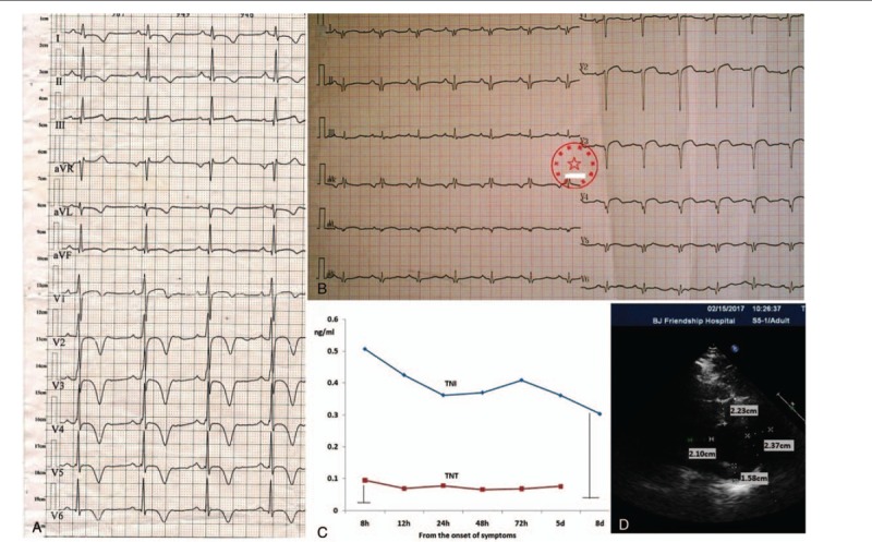
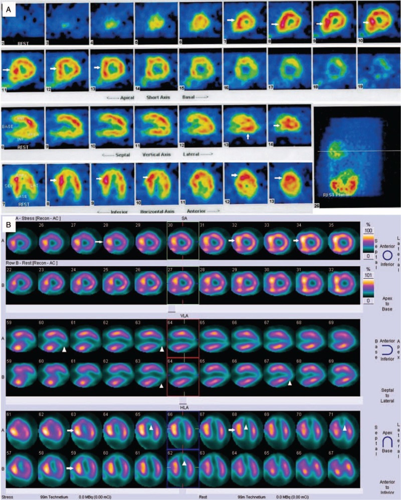
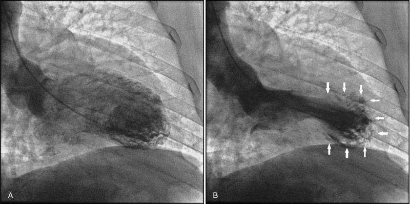
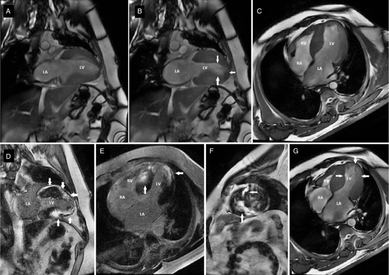
A 33-year-old man who presented with chest discomfort for 10 days. He had an emerging apical aneurysm in the LV without midventricular obstruction. He had been diagnosed with apical HCM via abnormal electrocardiograms (ECG) and single-photon emission computed tomography (SPECT) for 10 years. This time, a new significant change in ECG and SPECT was identified. Late gadolinium enhancement (LGE) was observed by cardiac magnetic resonance imaging (MRI), and SPECT showed myocardial fibrosis or necrosis involving the apical aneurysm and proximal portion of the heart, which was confirmed by left ventriculography.
We present a relatively rare case of HCM patients with apical aneurysms, accompaning by myocardial necrosis markers increased due to ventricular muscle stress increases, rather than obstructive coronary artery disease.
The patient was prescribed aspirin, metoprolol tartrate, perindopril, and atorvastatin and was strongly advised to quit cigarettes and reduce weight.
Follow-up at half a year turned out well.
LGE with a notable progression by ECG and SPECT along with an increase in myocardial necrosis markers in HCM patients with apical aneurysms, as was noted in the present case, is a relatively rare occurrence. Our present case may provide unique insights into the adverse remodelling process and the formation of apical aneurysms in HCM patients.
肥厚型心肌病(HCM)是一种以不适当的左心室和/或右心室肥厚及收缩亢进为特征的疾病,通常呈不对称性,且伴有心肌纤维排列紊乱的微观证据。本研究的目的是介绍一组此前未被充分认识的患有左心室心尖部动脉瘤的HCM患者。
一名33岁男性,因胸部不适10天前来就诊。他左心室出现了一个新的心尖部动脉瘤,无室中隔梗阻。他通过异常心电图(ECG)和单光子发射计算机断层扫描(SPECT)被诊断为心尖部HCM已有10年。此次,ECG和SPECT出现了新的显著变化。心脏磁共振成像(MRI)观察到晚期钆增强(LGE),SPECT显示心肌纤维化或坏死累及心尖部动脉瘤及心脏近端部分,左心室造影证实了这一点。
我们报告了一例相对罕见的患有心尖部动脉瘤的HCM患者,其心肌坏死标志物因心室肌压力增加而升高,而非梗阻性冠状动脉疾病所致。
给该患者开了阿司匹林、酒石酸美托洛尔、培哚普利和阿托伐他汀,并强烈建议其戒烟和减重。
半年随访情况良好。
如本病例所示,伴有心尖部动脉瘤的HCM患者中,LGE通过ECG和SPECT出现显著进展,同时心肌坏死标志物升高,这种情况相对罕见。我们目前的病例可能为HCM患者不良重塑过程及心尖部动脉瘤的形成提供独特见解。