Pneumology Research Group, Institut d'Investigació Biomèdica de Bellvitge - IDIBELL, L'Hospitalet de Llobregat, Barcelona, Spain.
Department of Respiratory Medicine, Unit of Chronic Obstructive Pulmonary Disease, Bellvitge University Hospital, L'Hospitalet de Llobregat, Barcelona, Spain.
Respir Res. 2018 May 28;19(1):103. doi: 10.1186/s12931-018-0793-0.
Extracellular adenosine triphosphate (ATP) is up-regulated in the airways of patients with chronic obstructive pulmonary disease (COPD), resulting in increased inflammation, bronchoconstriction, and cough. Although extracellular ATP levels are tightly controlled by nucleoside triphosphate diphosphohydrolase-1 (NTPDase1; also known as CD39) in the lungs, the role of CD39 in the pathology of COPD is unknown. We hypothesized that alterations in the expression and activity of CD39 could be part of the mechanisms for initiating and perpetuating the disease.
We analyzed CD39 gene and protein expression as well as ATPase enzyme activity in lung tissue samples of patients with COPD (n = 17), non-obstructed smokers (NOS) (n = 16), and never smokers (NS) (n = 13). Morphometry studies were performed to analyze pulmonary vascular remodeling.
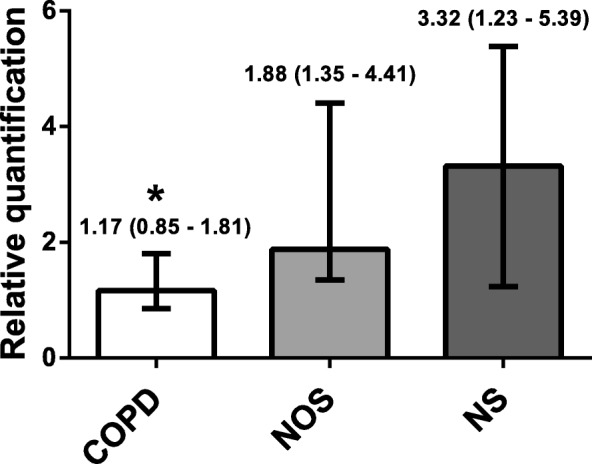
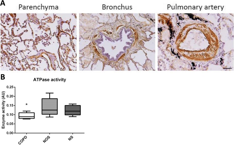
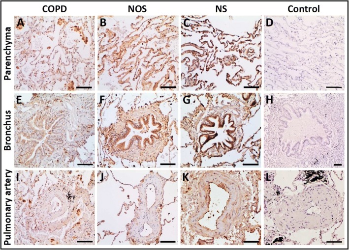
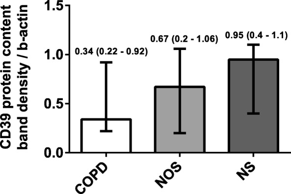
There was significantly decreased CD39 gene expression in the lungs of the COPD group (1.17 [0.85-1.81]) compared with the NOS group (1.88 [1.35-4.41]) and NS group (3.32 [1.23-5.39]) (p = 0.037). This attenuation correlated with higher systemic inflammation and intimal thickening of muscular pulmonary arteries in the COPD group. Lung CD39 protein levels were also lower in the COPD group (0.34 [0.22-0.92]) compared with the NOS group (0.67 [0.32-1.06]) and NS group (0.95 [0.4-1.1) (p = 0.133). Immunohistochemistry showed that CD39 was downregulated in lung parenchyma, epithelial bronchial cells, and the endothelial cells of pulmonary muscular arteries in the COPD group. ATPase activity in human pulmonary structures was reduced in the lungs of patients with COPD.
An attenuation of CD39 expression and activity is presented in lung tissue of stable COPD patients, which could lead to pulmonary ATP accumulation, favoring the development of pulmonary inflammation and emphysema. This may be a mechanism underlying the development of COPD.
细胞外三磷酸腺苷(ATP)在慢性阻塞性肺疾病(COPD)患者的气道中上调,导致炎症、支气管收缩和咳嗽增加。尽管细胞外 ATP 水平在肺部中受到核苷三磷酸二磷酸水解酶-1(NTPDase1;也称为 CD39)的严格控制,但 CD39 在 COPD 病理中的作用尚不清楚。我们假设 CD39 的表达和活性改变可能是引发和持续该疾病的机制之一。
我们分析了 COPD 患者(n=17)、非阻塞性吸烟者(NOS)(n=16)和从不吸烟者(NS)(n=13)的肺组织样本中的 CD39 基因和蛋白表达以及 ATP 酶活性。进行形态计量学研究以分析肺血管重塑。
与 NOS 组(1.88 [1.35-4.41])和 NS 组(3.32 [1.23-5.39])相比,COPD 组的肺 CD39 基因表达显著降低(1.17 [0.85-1.81])(p=0.037)。这种衰减与 COPD 组更高的全身炎症和肌性肺动脉内膜增厚相关。与 NOS 组(0.67 [0.32-1.06])和 NS 组(0.95 [0.4-1.1])相比,COPD 组的肺 CD39 蛋白水平也较低(0.34 [0.22-0.92])(p=0.133)。免疫组化显示,CD39 在 COPD 组的肺实质、上皮性支气管细胞和肺肌性动脉的内皮细胞中下调。COPD 患者的人肺结构中的 ATP 酶活性降低。
稳定的 COPD 患者的肺组织中 CD39 的表达和活性减弱,可能导致肺内 ATP 积累,有利于肺炎症和肺气肿的发展。这可能是 COPD 发展的一种机制。