Deyhimi Parviz, Alishahi Batoul
Dental Research Center, Dept. of Oral and Maxillofacial Pathology, School of Dentistry, Isfahan University of Medical Sciences, Isfahan, Iran.
Postgraduate Student, Dental Research Center, Dept. of Oral and Maxillofacial Pathology, School of Dentistry, Isfahan University of Medical Sciences, Isfahan, Iran.
J Dent (Shiraz). 2018 Jun;19(2):132-141.
Pemphigus vulgaris is characterized by intraepithelial vesicles, but pathogenesis of vesicle formation in this disease has not been substantiated yet.
The present study investigate extrinsic apoptotic pathway in oral pemphigus vulgaris using TUNEL and important immunohistochemical markers of extrinsic pathway, TNFR1 and FasL.
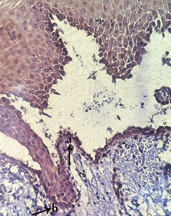
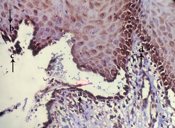
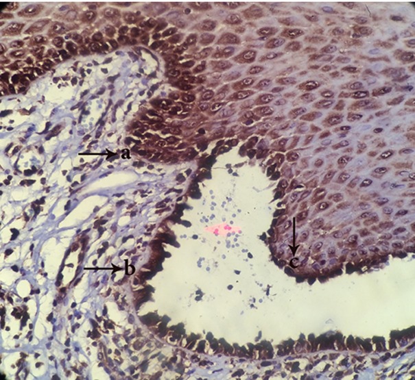
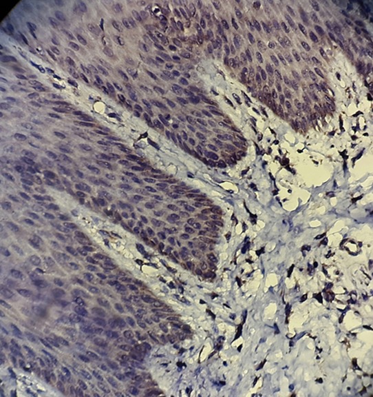
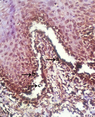
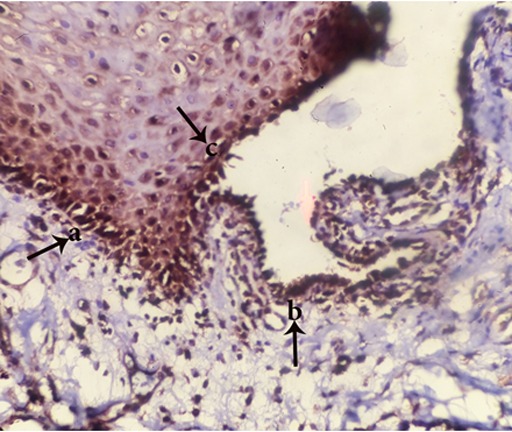
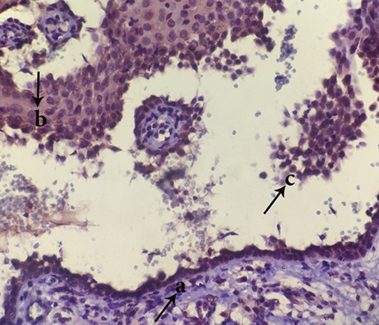
In the present cross sectional study, 25 oral pemphigus vulgaris samples and 6 normal oral mucosa were analyzed for the presence of apoptosis by TUNEL and the staining of TNFR1 and FasL in basal and parabasal layers around vesicle, vesicle floor, vesicle roof and acantholytic cells. The staining expression and intensity were measured and the obtained data were analyzed by Wilcoxon and Mann-Whitney tests.
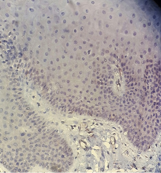
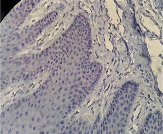
There was no or faint staining of TUNEL, FasL and TNFR1 in normal oral mucosa. In addition, there was no significant difference between the staining of TUNEL technique in different layers. The staining of TNFR1 marker was very high in all regions. FasL marker was not positive in the basal and parabasal layers around vesicle in 92% of samples but showed a varied and different staining in vesicle region. There was a significant difference between the each two markers in all layers ( <0.001).
Apoptosis is probably is a preceding phenomenon to acantholysis in pemphigus vulgaris. It appears that the apoptosis occurs mostly by extrinsic pathway using proapototic mediators TNFR1 and FasL.
寻常型天疱疮的特征为上皮内水疱,但该疾病中水疱形成的发病机制尚未得到证实。
本研究使用TUNEL以及外源性凋亡途径的重要免疫组织化学标志物TNFR1和FasL,研究口腔寻常型天疱疮中的外源性凋亡途径。
在本横断面研究中,通过TUNEL以及对水疱周围、水疱底部、水疱顶部和棘层松解细胞的基底层和副基底层中TNFR1和FasL进行染色,分析25例口腔寻常型天疱疮样本和6例正常口腔黏膜样本中凋亡的存在情况。测量染色表达和强度,并通过Wilcoxon检验和Mann-Whitney检验对所得数据进行分析。
正常口腔黏膜中TUNEL、FasL和TNFR1无染色或染色微弱。此外,不同层中TUNEL技术的染色之间无显著差异。TNFR1标志物在所有区域的染色都非常高。92%的样本中,水疱周围基底层和副基底层中的FasL标志物呈阴性,但在水疱区域显示出不同的染色情况。所有层中每两种标志物之间均存在显著差异(P<0.001)。
凋亡可能是寻常型天疱疮棘层松解之前的一种现象。似乎凋亡主要通过使用促凋亡介质TNFR1和FasL的外源性途径发生。