Department of Nephrology, The Second Xiangya Hospital, Central South University, Changsha, Hunan, China.
Changsha Central Hospital, Changsha, Hunan, China.
Oxid Med Cell Longev. 2018 May 16;2018:6296802. doi: 10.1155/2018/6296802. eCollection 2018.
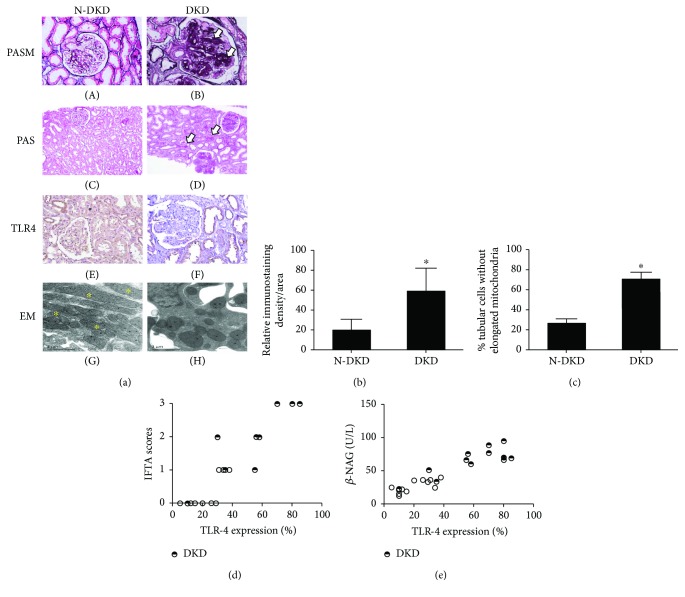
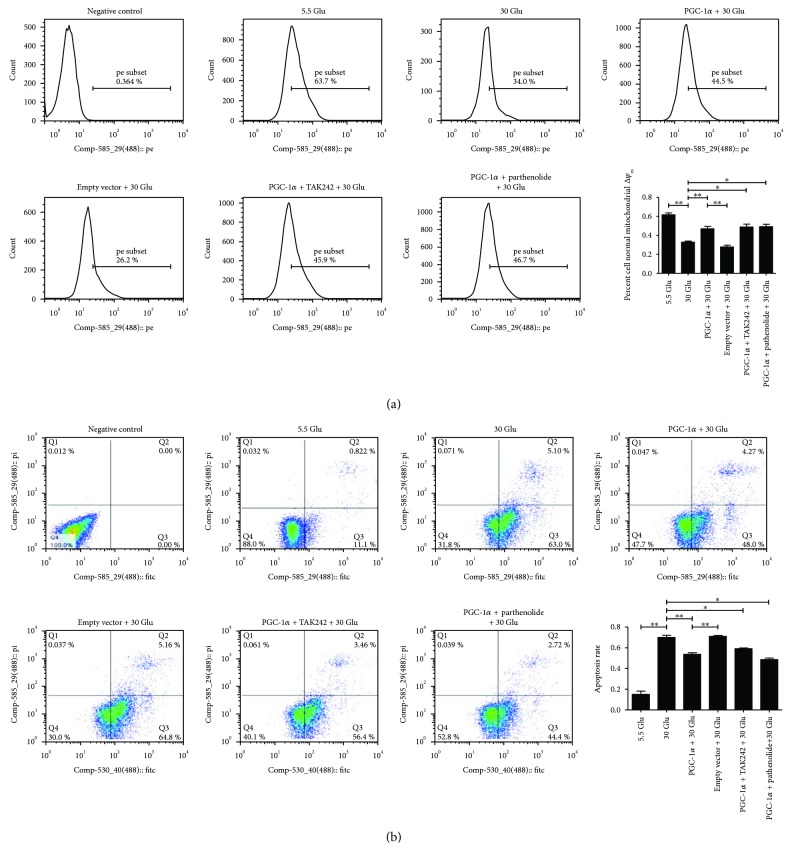
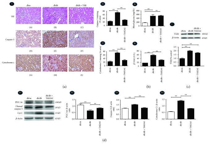
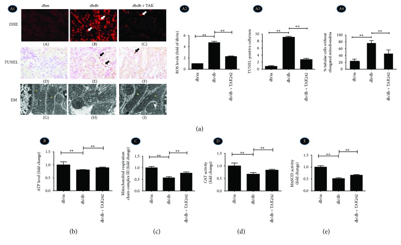
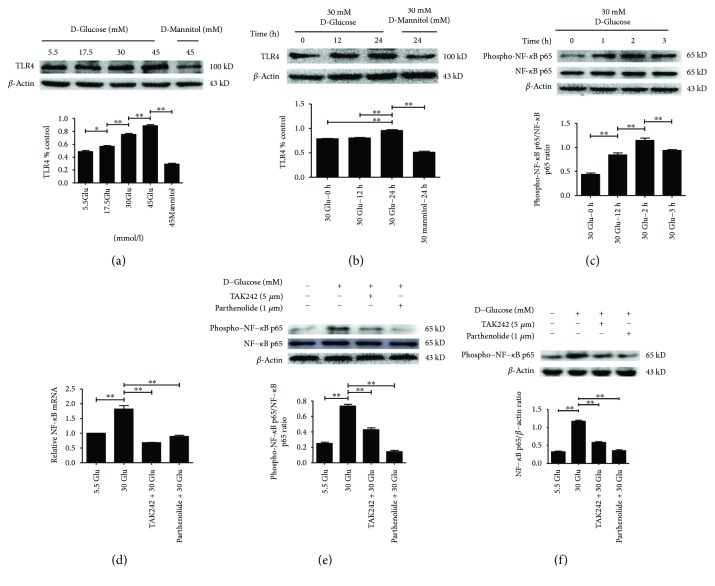
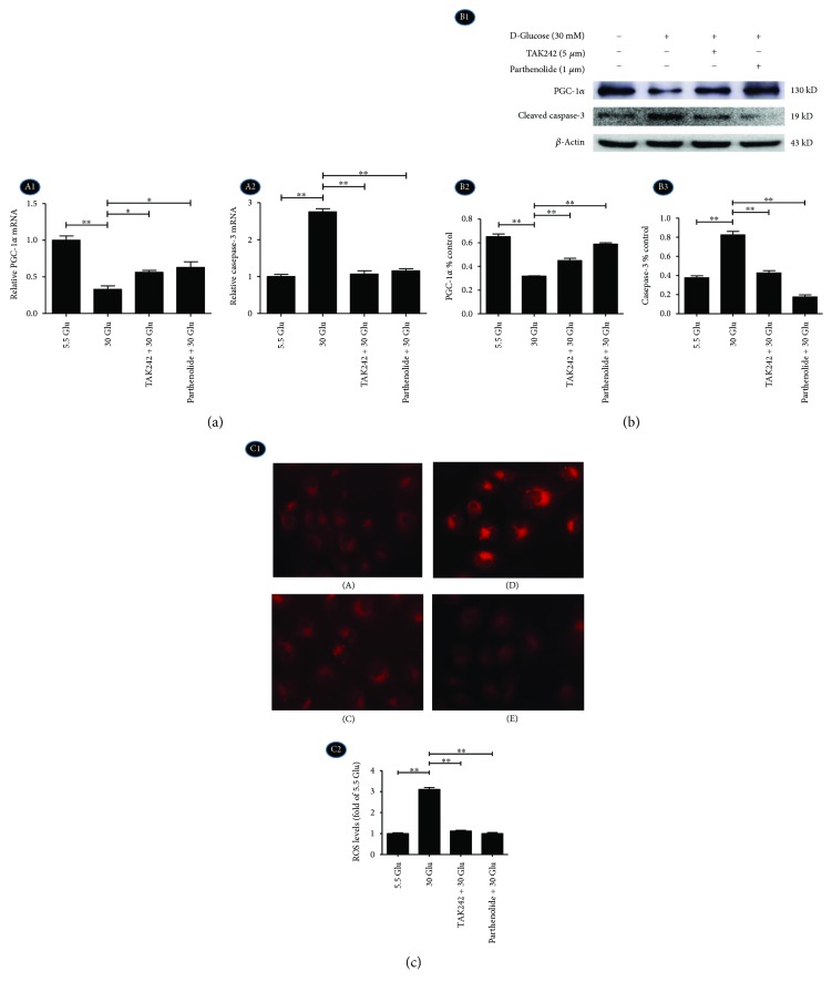
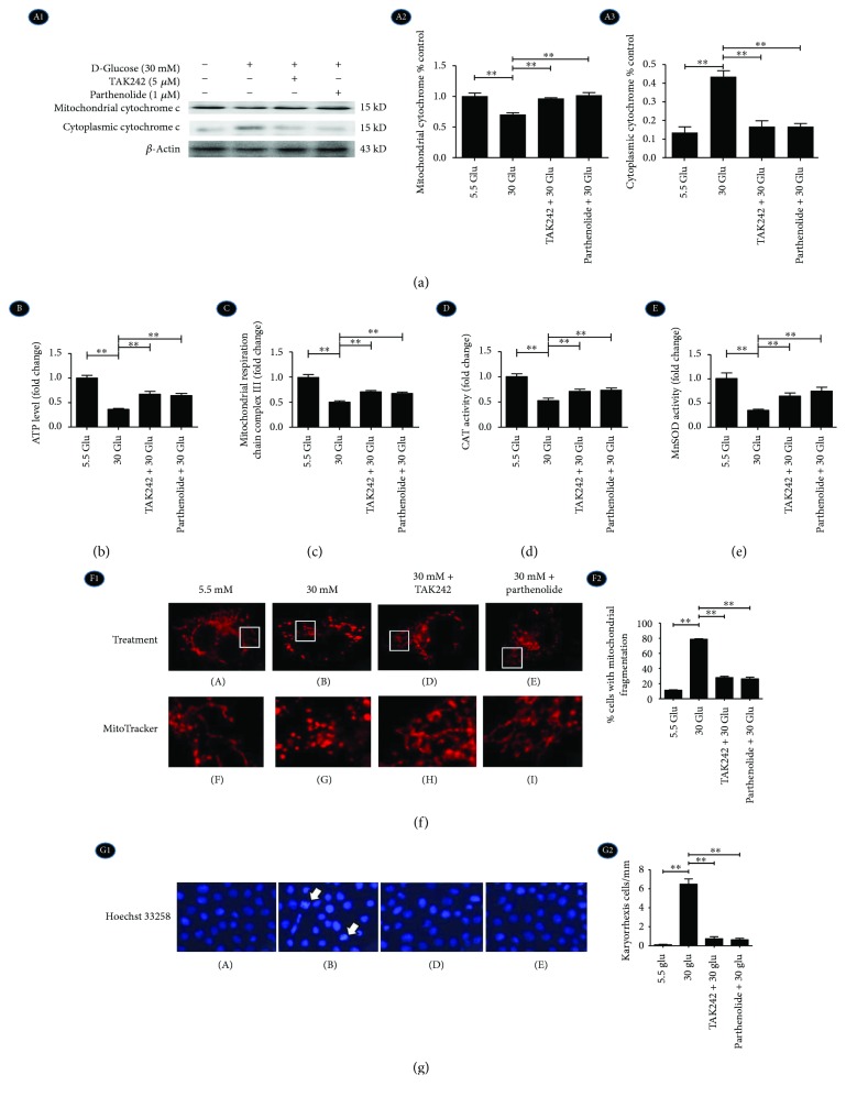
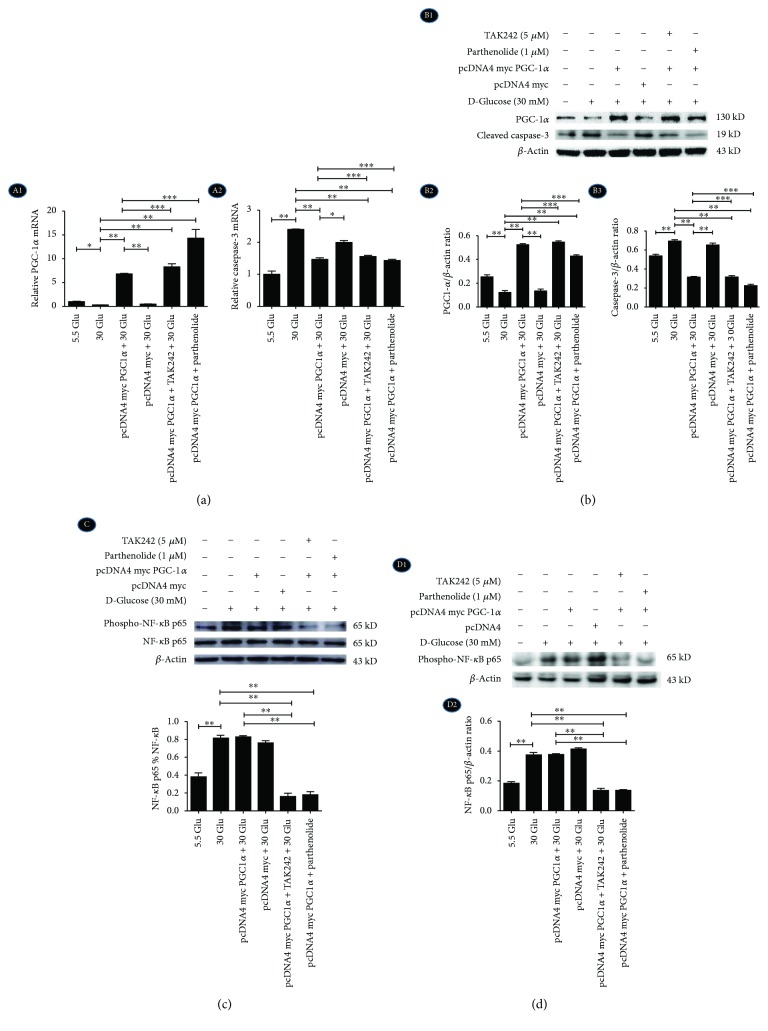
The role and precise mechanism of TLR4 in mitochondria-related oxidative damage and apoptosis of renal tubules in diabetic kidney disease (DKD) remain unclear. We examined the expression of TLR4 in renal biopsy tissues. Db/db diabetic mice and HK-2 cells cultured under high glucose (HG) were used as in vivo and vitro models. Real-time RT-PCR, Western blot, and immunohistochemistry were performed to examine the mRNA and protein levels of TLR4, NF-Β, PGC-1, cytochrome C, and cleaved caspase-3. ATP level, activity of electron transport chain complex III, and antioxidant enzymes were investigated for mitochondrial function. Electron microscopy (EM) and MitoTracker Red CMXRos were used for mitochondrial morphology alteration. DHE staining and TUNEL assay were detected for ROS accumulation and apoptosis. PGC-1 plasmids were used for the overexpression of PGC-1 in HK-2. TAK242 and parthenolide were used as TLR4 and NF-B blockers, respectively. Results showed that TLR4 was extensively expressed in the renal tubules of DKD patients and db/db diabetic mice, which was positively related to the tubular interstitial damage score and urinary -NAG levels. In diabetic mice, inhibition of TLR4 could reverse the decreased expression of PGC-1, increased expression of cytochrome C and cleaved caspase-3, mitochondrial dysfunction and deformation, increased accumulation of ROS, and activation of tubular cell apoptosis. In vitro, inhibition of TLR4 or NF-B showed consistent results. PGC-1 overexpression could reverse the mitochondrial dysfunction, increased cleaved caspase-3, and apoptosis in HK-2 cells treated with HG. Data indicated that the TLR4/NF-B signaling pathway might be the upstream pathway of PGC-1 and promote the tubular damage of DKD by modulating the mitochondria-related oxidative damage and apoptosis.
TLR4 在糖尿病肾病(DKD)肾小管线粒体相关氧化损伤和细胞凋亡中的作用及其确切机制尚不清楚。我们检测了 TLR4 在肾活检组织中的表达。Db/db 糖尿病小鼠和高糖(HG)培养的 HK-2 细胞分别作为体内和体外模型。采用实时 RT-PCR、Western blot 和免疫组化检测 TLR4、NF-Β、PGC-1、细胞色素 C 和 cleaved caspase-3 的 mRNA 和蛋白水平。检测线粒体功能的 ATP 水平、电子传递链复合物 III 活性和抗氧化酶。使用电子显微镜(EM)和 MitoTracker Red CMXRos 检测线粒体形态变化。DHE 染色和 TUNEL 检测 ROS 积累和细胞凋亡。使用 PGC-1 质粒过表达 HK-2 中的 PGC-1。TAK242 和白头翁内酯分别作为 TLR4 和 NF-B 的抑制剂。结果表明,TLR4 在 DKD 患者和 db/db 糖尿病小鼠的肾小管中广泛表达,与肾小管间质损伤评分和尿 -NAG 水平呈正相关。在糖尿病小鼠中,TLR4 抑制可逆转 PGC-1 表达降低、细胞色素 C 和 cleaved caspase-3 表达增加、线粒体功能障碍和变形、ROS 积累增加以及肾小管细胞凋亡激活。在体外,抑制 TLR4 或 NF-B 显示出一致的结果。PGC-1 过表达可逆转 HG 处理的 HK-2 细胞中线粒体功能障碍、增加的 cleaved caspase-3 和细胞凋亡。数据表明,TLR4/NF-B 信号通路可能是 PGC-1 的上游通路,通过调节与线粒体相关的氧化损伤和凋亡来促进 DKD 的肾小管损伤。