Zhang Can, Brandon Nicole R, Koper Kerryann, Tang Pei, Xu Yan, Dou Huanyu
1Departments of Anesthesiology.
2Pharmacology and Chemical Biology.
Aging Dis. 2018 Jun 1;9(3):412-425. doi: 10.14336/AD.2017.0926. eCollection 2018 Jun.
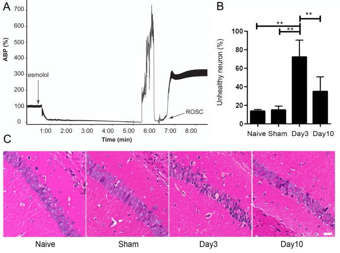
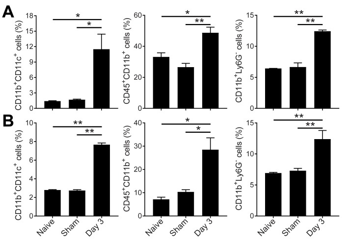
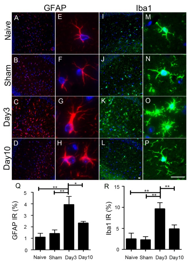
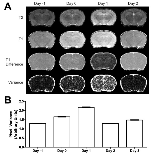
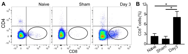
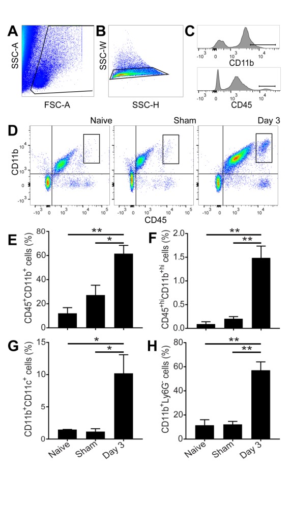
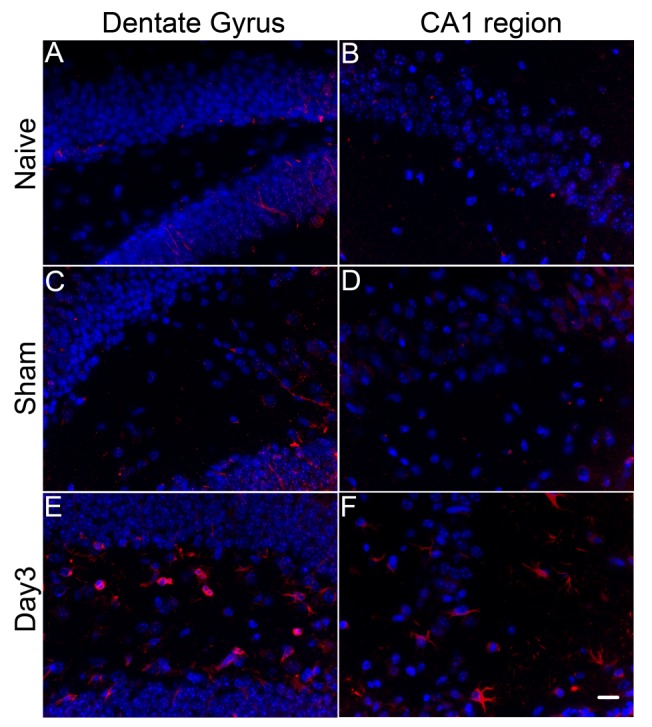
Although a direct link has long been suspected between systemic immune responses and neuronal injuries after stroke, it is unclear which immune cells play an important role. A question remains as to whether the blood brain barrier (BBB) is transiently disrupted after circulatory arrest to allow peripheral immune cells to enter brain parenchyma. Here, we developed a clinically relevant cardiac arrest and resuscitation model in mice to investigate the BBB integrity using noninvasive magnetic resonance imaging. Changes in immune signals in the brain and periphery were assayed by immunohistochemistry and flow cytometry. Quantitative variance maps from T1-weighted difference images before and after blood-pool contrast clearance revealed BBB disruptions immediately after resuscitation and one day after reperfusion. Time profiles of hippocampal CA1 neuronal injuries correlated with the morphological changes of microglia activation. Cytotoxic T cells, CD11bCD11c dendritic cells, and CD11bCD45 monocytes and macrophages were significantly increased in the brain three days after cardiac arrest and resuscitation, suggesting direct infiltration of these cells following the BBB disruption. Importantly, these immune cell changes were coupled with a parallel increase in the same subset of immune cell populations in the bone marrow and blood. We conclude that neurovascular breakdown during the initial reperfusion phase contributes to the systemic immune cell invasion and subsequent neuropathogenesis affecting the long-term outcome after cardiac arrest and resuscitation.
尽管长期以来人们一直怀疑全身免疫反应与中风后的神经元损伤之间存在直接联系,但尚不清楚哪些免疫细胞发挥重要作用。关于循环骤停后血脑屏障(BBB)是否会短暂破坏以允许外周免疫细胞进入脑实质,这一问题仍然存在。在此,我们在小鼠中建立了一个与临床相关的心脏骤停和复苏模型,以使用无创磁共振成像研究血脑屏障的完整性。通过免疫组织化学和流式细胞术检测脑和外周免疫信号的变化。血池造影剂清除前后T1加权差异图像的定量方差图显示,复苏后即刻和再灌注一天后血脑屏障被破坏。海马CA1神经元损伤的时间曲线与小胶质细胞激活的形态学变化相关。心脏骤停和复苏三天后,脑内细胞毒性T细胞、CD11bCD11c树突状细胞以及CD11bCD45单核细胞和巨噬细胞显著增加,表明血脑屏障破坏后这些细胞直接浸润。重要的是,这些免疫细胞变化与骨髓和血液中相同免疫细胞群体亚群的平行增加相关。我们得出结论,初始再灌注阶段的神经血管破坏导致全身免疫细胞入侵以及随后的神经病理发生,影响心脏骤停和复苏后的长期预后。